SHEET THE SHEET STRUCTURE OF THIS MOLECULE IS BIFURCATED. IN ORDER TO REPRESENT THIS FEATURE IN ... SHEET THE SHEET STRUCTURE OF THIS MOLECULE IS BIFURCATED. IN ORDER TO REPRESENT THIS FEATURE IN THE SHEET RECORDS BELOW, TWO SHEETS ARE DEFINED.
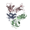
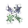
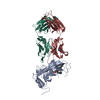
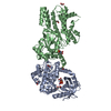
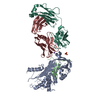
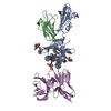
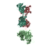
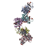
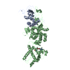
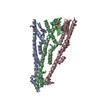
A: ABT-007 FAB FRAGMENT B: ERYTHROPOIETIN RECEPTOR C: ERYTHROPOIETIN RECEPTOR D: ABT-007 FAB FRAGMENT E: ERYTHROPOIETIN RECEPTOR F: ABT-007 FAB FRAGMENT G: ABT-007 FAB FRAGMENT H: ABT-007 FAB FRAGMENT L: ABT-007 FAB FRAGMENT
Mass: 23348.893 Da / Num. of mol.: 3 Source method: isolated from a genetically manipulated source Source: (gene. exp.) HOMO SAPIENS (human) / Production host: ESCHERICHIA COLI (E. coli) / References: UniProt: Q7Z3Y4*PLUS
#2: Protein
ERYTHROPOIETINRECEPTOR / / EPO-R
Mass: 24754.000 Da / Num. of mol.: 3 / Fragment: EPO RECEPTOR SOLUBLE DOMAIN, RESIDUES 25-249 Source method: isolated from a genetically manipulated source Source: (gene. exp.) HOMO SAPIENS (human) / Production host: ESCHERICHIA COLI (E. coli) / References: UniProt: P19235
#3: Antibody
ABT-007FABFRAGMENT
Mass: 22946.727 Da / Num. of mol.: 3 Source method: isolated from a genetically manipulated source Source: (gene. exp.) HOMO SAPIENS (human) / Production host: ESCHERICHIA COLI (E. coli) / References: UniProt: Q6GMX6*PLUS
-
Experimental details
-
Experiment
Experiment
Method: X-RAY DIFFRACTION / Number of used crystals: 1
-
Sample preparation
Crystal
Density Matthews: 3.55 Å3/Da / Density % sol: 65.33 % / Description: NONE
Protocol: SINGLE WAVELENGTH / Monochromatic (M) / Laue (L): M / Scattering type: x-ray
Radiation wavelength
Wavelength: 1 Å / Relative weight: 1
Reflection
Resolution: 3.2→50 Å / Num. obs: 50772 / % possible obs: 97.4 % / Observed criterion σ(I): 2 / Redundancy: 6.7 % / Rmerge(I) obs: 0.08 / Net I/σ(I): 15.4
Reflection shell
Resolution: 3.2→3.31 Å / Redundancy: 6 % / Rmerge(I) obs: 0.51 / Mean I/σ(I) obs: 2 / % possible all: 81.7
-
Processing
Software
Name
Version
Classification
HKL-2000
datareduction
SCALEPACK
datascaling
PHASER
phasing
MOLREP
phasing
REFMAC
5.2.0005
refinement
Refinement
Method to determine structure: MOLECULAR REPLACEMENT / Resolution: 3.2→113.23 Å / Cor.coef. Fo:Fc: 0.888 / Cor.coef. Fo:Fc free: 0.822 / SU B: 27.517 / SU ML: 0.475 / Cross valid method: THROUGHOUT / ESU R Free: 0.597 / Stereochemistry target values: MAXIMUM LIKELIHOOD / Details: HYDROGENS HAVE BEEN ADDED IN THE RIDING POSITIONS.
Rfactor
Num. reflection
% reflection
Selection details
Rfree
0.323
2506
5.1 %
RANDOM
Rwork
0.257
-
-
-
obs
0.26
46835
97.3 %
-
Solvent computation
Ion probe radii: 0.8 Å / Shrinkage radii: 0.8 Å / VDW probe radii: 1.2 Å / Solvent model: MASK
 Movie
Movie Controller

 Yorodumi
 Open data
 Basic information
 Components
 Keywords
 IMMUNE SYSTEM /
  Function and homology information
 Authors
 Citation
 Structure visualization
 Downloads & links
 Other downloads
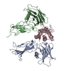


 PDBj
PDBj


 Assembly


 Sample preparation
 / Beamline: 17-ID / Wavelength: 1
  Processing