 Movie
Movie Controller

[English] 日本語
 Yorodumi
- PDB-1eba: COMPLEX BETWEEN THE EXTRACELLULAR DOMAIN OF ERYTHROPOIETIN (EPO) ... -
+ Open data

- Basic information
| Entry | Database: PDB / ID: 1eba | ||||||
|---|---|---|---|---|---|---|---|
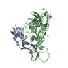
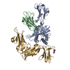
| Title | COMPLEX BETWEEN THE EXTRACELLULAR DOMAIN OF ERYTHROPOIETIN (EPO) RECEPTOR [EBP] AND AN INACTIVE PEPTIDE [EMP33] CONTAINS 3,5-DIBROMOTYROSINE IN POSITION 4 (DENOTED DBY) | ||||||
 Components Components |
| ||||||
 Keywords Keywords |  SIGNALING PROTEIN / ERYTHROPOIETIN RECEPTOR / SIGNAL TRANSDUCTION / PROTEIN MINIMIZATION / DRUG DESIGN / CYTOKINE RECEPTOR CLASS 1 / COMPLEX (CYTOKINE RECEPTOR-PEPTIDE) SIGNALING PROTEIN / ERYTHROPOIETIN RECEPTOR / SIGNAL TRANSDUCTION / PROTEIN MINIMIZATION / DRUG DESIGN / CYTOKINE RECEPTOR CLASS 1 / COMPLEX (CYTOKINE RECEPTOR-PEPTIDE) | ||||||
| Function / homology |  Function and homology informationerythropoietin receptor activity / Signaling by Erythropoietin / Erythropoietin activates STAT5 / Erythropoietin activates Phospholipase C gamma (PLCG) / Erythropoietin activates Phosphoinositide-3-kinase (PI3K) / hemopoiesis / decidualization / Erythropoietin activates RAS / brain development / cytokine-mediated signaling pathway ...erythropoietin receptor activity / Signaling by Erythropoietin / Erythropoietin activates STAT5 / Erythropoietin activates Phospholipase C gamma (PLCG) / Erythropoietin activates Phosphoinositide-3-kinase (PI3K) / hemopoiesis / decidualization / Erythropoietin activates RAS / brain development / cytokine-mediated signaling pathway / heart development / nuclear speck / external side of plasma membrane / positive regulation of cell population proliferation / signal transduction / extracellular region / identical protein binding / plasma membrane Function and homology informationerythropoietin receptor activity / Signaling by Erythropoietin / Erythropoietin activates STAT5 / Erythropoietin activates Phospholipase C gamma (PLCG) / Erythropoietin activates Phosphoinositide-3-kinase (PI3K) / hemopoiesis / decidualization / Erythropoietin activates RAS / brain development / cytokine-mediated signaling pathway ...erythropoietin receptor activity / Signaling by Erythropoietin / Erythropoietin activates STAT5 / Erythropoietin activates Phospholipase C gamma (PLCG) / Erythropoietin activates Phosphoinositide-3-kinase (PI3K) / hemopoiesis / decidualization / Erythropoietin activates RAS / brain development / cytokine-mediated signaling pathway / heart development / nuclear speck / external side of plasma membrane / positive regulation of cell population proliferation / signal transduction / extracellular region / identical protein binding / plasma membraneSimilarity search - Function | ||||||
| Biological species |  Homo sapiens (human) Homo sapiens (human) | ||||||
| Method | X-RAY DIFFRACTION / MOLECULAR REPLACEMENT / Resolution: 2.7 Å | ||||||
 Authors Authors | Livnah, O. / Stura, E.A. / Wilson, I.A. | ||||||
 Citation Citation | Journal: Nat.Struct.Biol. / Year: 1998 Title: An antagonist peptide-EPO receptor complex suggests that receptor dimerization is not sufficient for activation. Authors: Livnah, O. / Johnson, D.L. / Stura, E.A. / Farrell, F.X. / Barbone, F.P. / You, Y. / Liu, K.D. / Goldsmith, M.A. / He, W. / Krause, C.D. / Pestka, S. / Jolliffe, L.K. / Wilson, I.A. #1: Journal: Science / Year: 1996Title: Functional Mimicry of a Protein Hormone by a Peptide Agonist: The Epo Receptor Complex at 2.8 A Authors: Livnah, O. / Stura, E.A. / Johnson, D.L. / Middleton, S.A. / Mulcahy, L.S. / Wrighton, N.C. / Dower, W.J. / Jolliffe, L.K. / Wilson, I.A. | ||||||
| History |
|
- Structure visualization
| Structure viewer | Molecule: MolmilJmol/JSmol |
|---|
- Downloads & links
-Download
| PDBx/mmCIF format | 1eba.cif.gz | 98.1 KB | Display | PDBx/mmCIF format |
|---|---|---|---|---|
| PDB format | pdb1eba.ent.gz | 76 KB | Display | PDB format |
| PDBx/mmJSON format | 1eba.json.gz | Tree view | PDBx/mmJSON format | |
| Others |  Other downloads Other downloads |
-Validation report
| Arichive directory | https://data.pdbj.org/pub/pdb/validation_reports/eb/1ebaftp://data.pdbj.org/pub/pdb/validation_reports/eb/1eba | HTTPS FTP |
|---|
-Related structure data
| Related structure data |  1ebpS S: Starting model for refinement |
|---|---|
| Similar structure data |


-Links
 PDBj
PDBj


- Assembly
| Deposited unit | 
| ||||||||
|---|---|---|---|---|---|---|---|---|---|
| 1 |
| ||||||||
| 2 | 
| ||||||||
| 3 | 
| ||||||||
| Unit cell |
|
-Components
| #1: Protein | Mass: 23739.908 Da / Num. of mol.: 2 / Fragment: EXTRACELLULAR DOMAIN Source method: isolated from a genetically manipulated source Source: (gene. exp.) Homo sapiens (human) / Production host:  Escherichia coli (E. coli) / References: UniProt: P19235 Escherichia coli (E. coli) / References: UniProt: P19235#2: Protein/peptide | Mass: 2255.168 Da / Num. of mol.: 2 / Source method: obtained synthetically |
|---|
-Experimental details
-Experiment
| Experiment | Method: X-RAY DIFFRACTION / Number of used crystals: 1 |
|---|
- Sample preparation
| Crystal | Density Matthews: 2.77 Å3/Da / Density % sol: 55.61 % | |||||||||||||||
|---|---|---|---|---|---|---|---|---|---|---|---|---|---|---|---|---|
| Crystal grow | pH: 6 / Details: pH 6.0 | |||||||||||||||
| Crystal grow | *PLUS Temperature: 22 ℃ / pH: 6.5 / Method: vapor diffusion, sitting drop | |||||||||||||||
| Components of the solutions | *PLUS
|
-Data collection
| Diffraction | Mean temperature: 293 K |
|---|---|
| Diffraction source | Wavelength: 1.5418 |
| Detector | Type: MARRESEARCH / Detector: IMAGE PLATE / Details: MIRRORS |
| Radiation | Protocol: SINGLE WAVELENGTH / Monochromatic (M) / Laue (L): M / Scattering type: x-ray |
| Radiation wavelength | Wavelength: 1.5418 Å / Relative weight: 1 |
| Reflection | Resolution: 2.5→50 Å / Num. obs: 99999 / % possible obs: 99 % / Redundancy: 3.3 % / Rsym value: 0.089 / Net I/σ(I): 19.2 |
| Reflection shell | Resolution: 2.7→2.8 Å / Redundancy: 3.3 % / Mean I/σ(I) obs: 4.1 / Rsym value: 0.3 / % possible all: 99 |
| Reflection | *PLUS Lowest resolution: 50 Å / Num. obs: 16100 / Rmerge(I) obs: 0.089 |
| Reflection shell | *PLUS % possible obs: 99 % / Rmerge(I) obs: 0.3 |
- Processing
| Software |
| ||||||||||||||||||||||||||||||||||||||||||||||||||||||||||||
|---|---|---|---|---|---|---|---|---|---|---|---|---|---|---|---|---|---|---|---|---|---|---|---|---|---|---|---|---|---|---|---|---|---|---|---|---|---|---|---|---|---|---|---|---|---|---|---|---|---|---|---|---|---|---|---|---|---|---|---|---|---|
| Refinement | Method to determine structure: MOLECULAR REPLACEMENT Starting model: 1EBP MONOMER Resolution: 2.7→50 Å / Cross valid method: THROUGHT / σ(F): 1
| ||||||||||||||||||||||||||||||||||||||||||||||||||||||||||||
| Refinement step | Cycle: LAST / Resolution: 2.7→50 Å
| ||||||||||||||||||||||||||||||||||||||||||||||||||||||||||||
| Refine LS restraints |
| ||||||||||||||||||||||||||||||||||||||||||||||||||||||||||||
| Xplor file | Serial no: 1 / Param file: PARHCSDX | ||||||||||||||||||||||||||||||||||||||||||||||||||||||||||||
| Software | *PLUS Name: X-PLOR / Version: 3.8 / Classification: refinement | ||||||||||||||||||||||||||||||||||||||||||||||||||||||||||||
| Refinement | *PLUS Highest resolution: 2.7 Å / Lowest resolution: 50 Å / σ(F): 1 / Num. reflection Rfree: 1498 / % reflection Rfree: 10 % / Rfactor obs: 0.2 / Rfactor Rwork: 0.2 | ||||||||||||||||||||||||||||||||||||||||||||||||||||||||||||
| Solvent computation | *PLUS | ||||||||||||||||||||||||||||||||||||||||||||||||||||||||||||
| Displacement parameters | *PLUS | ||||||||||||||||||||||||||||||||||||||||||||||||||||||||||||
| Refine LS restraints | *PLUS
|
