 Movie
Movie Controller


+ Open data

- Basic information
| Entry | Database: PDB / ID: 5y3c | ||||||||||||||||||||||||||||||||||||||||||||||||||||||
|---|---|---|---|---|---|---|---|---|---|---|---|---|---|---|---|---|---|---|---|---|---|---|---|---|---|---|---|---|---|---|---|---|---|---|---|---|---|---|---|---|---|---|---|---|---|---|---|---|---|---|---|---|---|---|---|
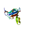
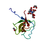
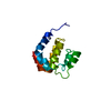
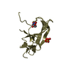
| Title | Crystal structure of zebrafish Ccd1 DIX domain | ||||||||||||||||||||||||||||||||||||||||||||||||||||||
 Components Components | Dixin-A | ||||||||||||||||||||||||||||||||||||||||||||||||||||||
 Keywords Keywords |  SIGNALING PROTEIN / Wnt signal SIGNALING PROTEIN / Wnt signal | ||||||||||||||||||||||||||||||||||||||||||||||||||||||
| Function / homology |  Function and homology information Function and homology informationcamera-type eye development / canonical Wnt signaling pathway / forebrain development / focal adhesion / identical protein binding / cytosolSimilarity search - Function | ||||||||||||||||||||||||||||||||||||||||||||||||||||||
| Biological species |  Danio rerio (zebrafish) Danio rerio (zebrafish) | ||||||||||||||||||||||||||||||||||||||||||||||||||||||
| Method | X-RAY DIFFRACTION / SYNCHROTRON / MOLECULAR REPLACEMENT / Resolution: 1.96 Å | ||||||||||||||||||||||||||||||||||||||||||||||||||||||
 Authors Authors | Terawaki, S. / Shibata, N. / Higuchi, Y. | ||||||||||||||||||||||||||||||||||||||||||||||||||||||
| Funding support |  Japan, 17items Japan, 17items
| ||||||||||||||||||||||||||||||||||||||||||||||||||||||
 Citation Citation | Journal: Sci Rep / Year: 2017 Title: Structural basis for Ccd1 auto-inhibition in the Wnt pathway through homomerization of the DIX domain. Authors: Terawaki, S.I. / Fujita, S. / Katsutani, T. / Shiomi, K. / Keino-Masu, K. / Masu, M. / Wakamatsu, K. / Shibata, N. / Higuchi, Y. #1: Journal: Nat. Struct. Mol. Biol. / Year: 2007 Title: The DIX domain of Dishevelled confers Wnt signaling by dynamic polymerization. Authors: Schwarz-Romond, T. / Fiedler, M. / Shibata, N. / Butler, P.J. / Kikuchi, A. / Higuchi, Y. / Bienz, M. | ||||||||||||||||||||||||||||||||||||||||||||||||||||||
| History |
|
- Structure visualization
| Structure viewer | Molecule: MolmilJmol/JSmol |
|---|
- Downloads & links
-Download
| PDBx/mmCIF format | 5y3c.cif.gz | 51.1 KB | Display | PDBx/mmCIF format |
|---|---|---|---|---|
| PDB format | pdb5y3c.ent.gz | 35.5 KB | Display | PDB format |
| PDBx/mmJSON format | 5y3c.json.gz | Tree view | PDBx/mmJSON format | |
| Others |  Other downloads Other downloads |
-Validation report
| Arichive directory | https://data.pdbj.org/pub/pdb/validation_reports/y3/5y3cftp://data.pdbj.org/pub/pdb/validation_reports/y3/5y3c | HTTPS FTP |
|---|
-Related structure data
| Related structure data | 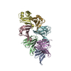 5y3bSC S: Starting model for refinement C: citing same article ( |
|---|---|
| Similar structure data |


-Links
 PDBj
PDBj- Assembly
| Deposited unit | 
| ||||||||
|---|---|---|---|---|---|---|---|---|---|
| 1 |
| ||||||||
| Unit cell |
|
-Components
| #1: Protein | Mass: 9598.747 Da / Num. of mol.: 1 / Fragment: UNP RESIDUES 356-438 Source method: isolated from a genetically manipulated source Source: (gene. exp.) Danio rerio (zebrafish) / Gene: dixdc1a, ccd1, dixdc1 / Plasmid: pOPINM / Production host:  Escherichia coli (E. coli) / Strain (production host): BL21(DE3)star / References: UniProt: Q804T6 Escherichia coli (E. coli) / Strain (production host): BL21(DE3)star / References: UniProt: Q804T6 |
|---|---|
| #2: Water | ChemComp-HOH / Water Mass: 18.015 Da / Num. of mol.: 66 / Source method: isolated from a natural source / Formula: H2O Mass: 18.015 Da / Num. of mol.: 66 / Source method: isolated from a natural source / Formula: H2O |
-Experimental details
-Experiment
| Experiment | Method: X-RAY DIFFRACTION / Number of used crystals: 1 |
|---|
- Sample preparation
| Crystal | Density Matthews: 2.61 Å3/Da / Density % sol: 52.79 % |
|---|---|
| Crystal grow | Temperature: 283 K / Method: vapor diffusion, sitting drop / pH: 7.5 Details: 14% poly(acrylic acid sodium salt) 5100, 100 mM HEPES-Na (pH 7.5), 20 mM MgCl2, 10 mM TCEP-HCl |
-Data collection
| Diffraction | Mean temperature: 100 K |
|---|---|
| Diffraction source | Source: SYNCHROTRON / Site: Photon Factory / Beamline: BL-1A / Wavelength: 1.1 Å |
| Detector | Type: DECTRIS PILATUS 2M-F / Detector: PIXEL / Date: Dec 21, 2014 |
| Radiation | Protocol: SINGLE WAVELENGTH / Monochromatic (M) / Laue (L): M / Scattering type: x-ray |
| Radiation wavelength | Wavelength: 1.1 Å / Relative weight: 1 |
| Reflection | Resolution: 1.96→50 Å / Num. obs: 7076 / % possible obs: 99.1 % / Redundancy: 9 % / Net I/σ(I): 35.3 |
- Processing
| Software |
| ||||||||||||||||||||||||||||||||||||||||||||||||||||||||||||||||||||||||||||||||||||||||||||||||||||||||||||||||||||||||||||||||||||||||||||||||||||||||||||||||||||||||||||||||||||||||||||||||||||||||||||||||||||||||||||||||||||||||||||||||||||||||||||||||||||||||||||||||||||||||||||||||||||||||||||
|---|---|---|---|---|---|---|---|---|---|---|---|---|---|---|---|---|---|---|---|---|---|---|---|---|---|---|---|---|---|---|---|---|---|---|---|---|---|---|---|---|---|---|---|---|---|---|---|---|---|---|---|---|---|---|---|---|---|---|---|---|---|---|---|---|---|---|---|---|---|---|---|---|---|---|---|---|---|---|---|---|---|---|---|---|---|---|---|---|---|---|---|---|---|---|---|---|---|---|---|---|---|---|---|---|---|---|---|---|---|---|---|---|---|---|---|---|---|---|---|---|---|---|---|---|---|---|---|---|---|---|---|---|---|---|---|---|---|---|---|---|---|---|---|---|---|---|---|---|---|---|---|---|---|---|---|---|---|---|---|---|---|---|---|---|---|---|---|---|---|---|---|---|---|---|---|---|---|---|---|---|---|---|---|---|---|---|---|---|---|---|---|---|---|---|---|---|---|---|---|---|---|---|---|---|---|---|---|---|---|---|---|---|---|---|---|---|---|---|---|---|---|---|---|---|---|---|---|---|---|---|---|---|---|---|---|---|---|---|---|---|---|---|---|---|---|---|---|---|---|---|---|---|---|---|---|---|---|---|---|---|---|---|---|---|---|---|---|---|---|---|---|---|---|---|---|---|---|---|---|---|---|---|---|---|---|---|---|---|---|---|---|---|---|---|---|---|---|---|---|---|---|
| Refinement | Method to determine structure: MOLECULAR REPLACEMENT Starting model: 5Y3B Resolution: 1.96→23.362 Å / SU ML: 0.18 / Cross valid method: FREE R-VALUE / σ(F): 1.38 / Phase error: 22.06
| ||||||||||||||||||||||||||||||||||||||||||||||||||||||||||||||||||||||||||||||||||||||||||||||||||||||||||||||||||||||||||||||||||||||||||||||||||||||||||||||||||||||||||||||||||||||||||||||||||||||||||||||||||||||||||||||||||||||||||||||||||||||||||||||||||||||||||||||||||||||||||||||||||||||||||||
| Solvent computation | Shrinkage radii: 0.9 Å / VDW probe radii: 1.11 Å | ||||||||||||||||||||||||||||||||||||||||||||||||||||||||||||||||||||||||||||||||||||||||||||||||||||||||||||||||||||||||||||||||||||||||||||||||||||||||||||||||||||||||||||||||||||||||||||||||||||||||||||||||||||||||||||||||||||||||||||||||||||||||||||||||||||||||||||||||||||||||||||||||||||||||||||
| Refinement step | Cycle: LAST / Resolution: 1.96→23.362 Å
| ||||||||||||||||||||||||||||||||||||||||||||||||||||||||||||||||||||||||||||||||||||||||||||||||||||||||||||||||||||||||||||||||||||||||||||||||||||||||||||||||||||||||||||||||||||||||||||||||||||||||||||||||||||||||||||||||||||||||||||||||||||||||||||||||||||||||||||||||||||||||||||||||||||||||||||
| Refine LS restraints |
| ||||||||||||||||||||||||||||||||||||||||||||||||||||||||||||||||||||||||||||||||||||||||||||||||||||||||||||||||||||||||||||||||||||||||||||||||||||||||||||||||||||||||||||||||||||||||||||||||||||||||||||||||||||||||||||||||||||||||||||||||||||||||||||||||||||||||||||||||||||||||||||||||||||||||||||
| LS refinement shell |
| ||||||||||||||||||||||||||||||||||||||||||||||||||||||||||||||||||||||||||||||||||||||||||||||||||||||||||||||||||||||||||||||||||||||||||||||||||||||||||||||||||||||||||||||||||||||||||||||||||||||||||||||||||||||||||||||||||||||||||||||||||||||||||||||||||||||||||||||||||||||||||||||||||||||||||||
| Refinement TLS params. | Method: refined / Refine-ID: X-RAY DIFFRACTION
| ||||||||||||||||||||||||||||||||||||||||||||||||||||||||||||||||||||||||||||||||||||||||||||||||||||||||||||||||||||||||||||||||||||||||||||||||||||||||||||||||||||||||||||||||||||||||||||||||||||||||||||||||||||||||||||||||||||||||||||||||||||||||||||||||||||||||||||||||||||||||||||||||||||||||||||
| Refinement TLS group |
|
