| Entry | Database: PDB / ID: 6f55
|
|---|
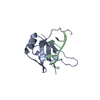
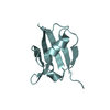
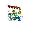
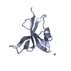
| Title | Complex structure of PACSIN SH3 domain and TRPV4 proline rich region |
|---|
 Components Components | |
|---|
 Keywords Keywords | PEPTIDE BINDING PROTEIN /  PROTEIN PROTEIN |
|---|
| Function / homology |  Function and homology information Function and homology information |
|---|
| Biological species |   Gallus gallus (chicken) Gallus gallus (chicken)
 Homo sapiens (human) Homo sapiens (human) |
|---|
| Method | SOLUTION NMR / torsion angle dynamics |
|---|
 Authors Authors | Glogowski, N.A. / Goretzki, B. / Diehl, E. / Duchardt-Ferner, E. / Hacker, C. / Hellmich, U.A. |
|---|
 Citation Citation | Journal: Structure / Year: 2018
Title: Structural Basis of TRPV4 N Terminus Interaction with Syndapin/PACSIN1-3 and PIP2.
Authors: Goretzki, B. / Glogowski, N.A. / Diehl, E. / Duchardt-Ferner, E. / Hacker, C. / Gaudet, R. / Hellmich, U.A. |
|---|
| History | | Deposition | Nov 30, 2017 | Deposition site: PDBE / Processing site: PDBE |
|---|
| Revision 1.0 | Oct 3, 2018 | Provider: repository / Type: Initial release |
|---|
| Revision 1.1 | Dec 19, 2018 | Group: Data collection / Database references / Category: citation
Item: _citation.journal_volume / _citation.page_first / _citation.page_last |
|---|
| Revision 1.2 | May 8, 2019 | Group: Data collection / Category: pdbx_nmr_software / Item: _pdbx_nmr_software.name |
|---|
| Revision 1.3 | May 15, 2024 | Group: Data collection / Database references
Category: chem_comp_atom / chem_comp_bond ...chem_comp_atom / chem_comp_bond / database_2 / pdbx_nmr_spectrometer
Item: _database_2.pdbx_DOI / _database_2.pdbx_database_accession / _pdbx_nmr_spectrometer.model |
|---|
|
|---|
 Movie
Movie Controller

 Yorodumi
 Open data
 Basic information
 Structure visualization
 Downloads & links
 Other downloads
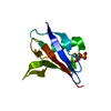


 PDBj
PDBj


 Assembly

 Sample preparation