 Movie
Movie Controller


+ Open data

- Basic information
| Entry | Database: PDB / ID: 5y3b | ||||||||||||||||||||||||||||||||||||||||||||||||||||||
|---|---|---|---|---|---|---|---|---|---|---|---|---|---|---|---|---|---|---|---|---|---|---|---|---|---|---|---|---|---|---|---|---|---|---|---|---|---|---|---|---|---|---|---|---|---|---|---|---|---|---|---|---|---|---|---|
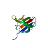
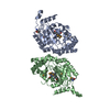
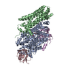
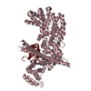
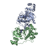
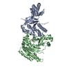
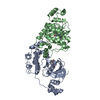
| Title | Crystal structure of mouse Ccd1 DIX domain | ||||||||||||||||||||||||||||||||||||||||||||||||||||||
 Components Components | Dixin | ||||||||||||||||||||||||||||||||||||||||||||||||||||||
 Keywords Keywords |  SIGNALING PROTEIN / Wnt signal SIGNALING PROTEIN / Wnt signal | ||||||||||||||||||||||||||||||||||||||||||||||||||||||
| Function / homology |  Function and homology information Function and homology informationforebrain ventricular zone progenitor cell division / cerebral cortex radially oriented cell migration / cell proliferation in forebrain / cerebellar cortex development / mitogen-activated protein kinase kinase kinase binding / gamma-tubulin binding / positive regulation of axonogenesis / cerebral cortex cell migration / positive regulation of Wnt signaling pathway / negative regulation of neuron differentiation ...forebrain ventricular zone progenitor cell division / cerebral cortex radially oriented cell migration / cell proliferation in forebrain / cerebellar cortex development / mitogen-activated protein kinase kinase kinase binding / gamma-tubulin binding / positive regulation of axonogenesis / cerebral cortex cell migration / positive regulation of Wnt signaling pathway / negative regulation of neuron differentiation / canonical Wnt signaling pathway / axon terminus / regulation of microtubule cytoskeleton organization / regulation of actin cytoskeleton organization / positive regulation of JNK cascade / actin binding / cytoskeleton / protein domain specific binding / focal adhesion / neuronal cell body / protein-containing complex / cytosol / cytoplasmSimilarity search - Function | ||||||||||||||||||||||||||||||||||||||||||||||||||||||
| Biological species |  Mus musculus (house mouse) Mus musculus (house mouse) | ||||||||||||||||||||||||||||||||||||||||||||||||||||||
| Method | X-RAY DIFFRACTION / SYNCHROTRON / MAD / Resolution: 3 Å | ||||||||||||||||||||||||||||||||||||||||||||||||||||||
 Authors Authors | Terawaki, S. / Shibata, N. / Higuchi, Y. | ||||||||||||||||||||||||||||||||||||||||||||||||||||||
| Funding support |  Japan, 17items Japan, 17items
| ||||||||||||||||||||||||||||||||||||||||||||||||||||||
 Citation Citation | Journal: Sci Rep / Year: 2017 Title: Structural basis for Ccd1 auto-inhibition in the Wnt pathway through homomerization of the DIX domain. Authors: Terawaki, S.I. / Fujita, S. / Katsutani, T. / Shiomi, K. / Keino-Masu, K. / Masu, M. / Wakamatsu, K. / Shibata, N. / Higuchi, Y. #1: Journal: Nat. Struct. Mol. Biol. / Year: 2007 Title: The DIX domain of Dishevelled confers Wnt signaling by dynamic polymerization. Authors: Schwarz-Romond, T. / Fiedler, M. / Shibata, N. / Butler, P.J. / Kikuchi, A. / Higuchi, Y. / Bienz, M. | ||||||||||||||||||||||||||||||||||||||||||||||||||||||
| History |
|
- Structure visualization
| Structure viewer | Molecule: MolmilJmol/JSmol |
|---|
- Downloads & links
-Download
| PDBx/mmCIF format | 5y3b.cif.gz | 125.4 KB | Display | PDBx/mmCIF format |
|---|---|---|---|---|
| PDB format | pdb5y3b.ent.gz | 99.1 KB | Display | PDB format |
| PDBx/mmJSON format | 5y3b.json.gz | Tree view | PDBx/mmJSON format | |
| Others |  Other downloads Other downloads |
-Validation report
| Arichive directory | https://data.pdbj.org/pub/pdb/validation_reports/y3/5y3bftp://data.pdbj.org/pub/pdb/validation_reports/y3/5y3b | HTTPS FTP |
|---|
-Related structure data


-Links
 PDBj
PDBj- Assembly
| Deposited unit | 
| ||||||||||||||||||||||||||||||||||||||||||||||||
|---|---|---|---|---|---|---|---|---|---|---|---|---|---|---|---|---|---|---|---|---|---|---|---|---|---|---|---|---|---|---|---|---|---|---|---|---|---|---|---|---|---|---|---|---|---|---|---|---|---|
| 1 |
| ||||||||||||||||||||||||||||||||||||||||||||||||
| Unit cell |
| ||||||||||||||||||||||||||||||||||||||||||||||||
| Noncrystallographic symmetry (NCS) | NCS domain:
NCS domain segments:
|
-Components
| #1: Protein | Mass: 9783.971 Da / Num. of mol.: 7 / Fragment: UNP RESIDUES 625-707 Source method: isolated from a genetically manipulated source Source: (gene. exp.) Mus musculus (house mouse) / Gene: Dixdc1, Ccd1, Kiaa1735 / Plasmid: pET49b / Production host:  Escherichia coli (E. coli) / Strain (production host): BL21 (DE3)-CodonPlus-RILP / References: UniProt: Q80Y83 Escherichia coli (E. coli) / Strain (production host): BL21 (DE3)-CodonPlus-RILP / References: UniProt: Q80Y83 |
|---|
-Experimental details
-Experiment
| Experiment | Method: X-RAY DIFFRACTION / Number of used crystals: 1 |
|---|
- Sample preparation
| Crystal | Density Matthews: 2.53 Å3/Da / Density % sol: 51.33 % |
|---|---|
| Crystal grow | Temperature: 293 K / Method: vapor diffusion, hanging drop / pH: 7.8 Details: 0.1 M Na HEPES pH 7.8, 15%(v/v) ethylene glycol, 3%(v/v) glycerol, 4%(v/v) 1,3-propanediol |
-Data collection
| Diffraction | Mean temperature: 100 K |
|---|---|
| Diffraction source | Source: SYNCHROTRON / Site: SPring-8 / Beamline: BL26B2 / Wavelength: 1 Å |
| Detector | Type: ADSC QUANTUM 315 / Detector: CCD / Date: Feb 20, 2009 |
| Radiation | Protocol: MAD / Monochromatic (M) / Laue (L): M / Scattering type: x-ray |
| Radiation wavelength | Wavelength: 1 Å / Relative weight: 1 |
| Reflection | Resolution: 3→50 Å / Num. obs: 14312 / % possible obs: 99 % / Redundancy: 6.2 % / Net I/σ(I): 16.3 |
- Processing
| Software |
| ||||||||||||||||||||||||||||||||||||||||||||||||||||||||
|---|---|---|---|---|---|---|---|---|---|---|---|---|---|---|---|---|---|---|---|---|---|---|---|---|---|---|---|---|---|---|---|---|---|---|---|---|---|---|---|---|---|---|---|---|---|---|---|---|---|---|---|---|---|---|---|---|---|
| Refinement | Method to determine structure: MAD / Resolution: 3→29.088 Å / SU ML: 0.42 / Cross valid method: FREE R-VALUE / σ(F): 1.34 / Phase error: 29.51
| ||||||||||||||||||||||||||||||||||||||||||||||||||||||||
| Solvent computation | Shrinkage radii: 0.9 Å / VDW probe radii: 1.11 Å / Bsol: 27.027 Å2 / ksol: 0.29 e/Å3 | ||||||||||||||||||||||||||||||||||||||||||||||||||||||||
| Displacement parameters |
| ||||||||||||||||||||||||||||||||||||||||||||||||||||||||
| Refinement step | Cycle: LAST / Resolution: 3→29.088 Å
| ||||||||||||||||||||||||||||||||||||||||||||||||||||||||
| Refine LS restraints |
| ||||||||||||||||||||||||||||||||||||||||||||||||||||||||
| Refine LS restraints NCS |
| ||||||||||||||||||||||||||||||||||||||||||||||||||||||||
| LS refinement shell |
|
