 Movie
Movie Controller

[English] 日本語
 Yorodumi
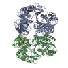
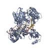
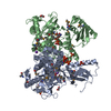
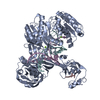
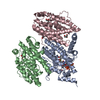
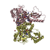
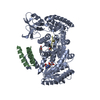
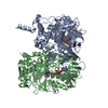
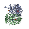
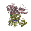
- PDB-5xpg: Crystal structure of T. thermophilus Argonaute protein complexed ... -
+ Open data

- Basic information
| Entry | Database: PDB / ID: 5xpg | ||||||
|---|---|---|---|---|---|---|---|
| Title | Crystal structure of T. thermophilus Argonaute protein complexed with a bulge 6'U7' on the target strand | ||||||
 Components Components |
| ||||||
 Keywords Keywords |  DNA BINDING PROTEIN / Argonaute / miRNA / bulge / mismatch DNA BINDING PROTEIN / Argonaute / miRNA / bulge / mismatch | ||||||
| Function / homology |  Function and homology informationHydrolases; Acting on ester bonds; Site specific endodeoxyribonucleases: cleavage is not sequence specific (deleted sub-subclass) / clearance of foreign intracellular DNA / DNA endonuclease activity / manganese ion binding / DNA replication / DNA binding / RNA binding Function and homology informationHydrolases; Acting on ester bonds; Site specific endodeoxyribonucleases: cleavage is not sequence specific (deleted sub-subclass) / clearance of foreign intracellular DNA / DNA endonuclease activity / manganese ion binding / DNA replication / DNA binding / RNA bindingSimilarity search - Function | ||||||
| Biological species |   Thermus thermophilus (bacteria) Thermus thermophilus (bacteria) | ||||||
| Method | X-RAY DIFFRACTION / SYNCHROTRON / MOLECULAR REPLACEMENT / Resolution: 2.8 Å | ||||||
 Authors Authors | Sheng, G. / Gogakos, T. / Wang, J. / Zhao, H. / Serganov, A. / Juranek, S. / Tuschl, T. / Patel, J.D. / Wang, Y. | ||||||
 Citation Citation | Journal: Nucleic Acids Res. / Year: 2017 Title: Structure/cleavage-based insights into helical perturbations at bulge sites within T. thermophilus Argonaute silencing complexes. Authors: Sheng, G. / Gogakos, T. / Wang, J. / Zhao, H. / Serganov, A. / Juranek, S. / Tuschl, T. / Patel, D.J. / Wang, Y. | ||||||
| History |
|
- Structure visualization
| Structure viewer | Molecule: MolmilJmol/JSmol |
|---|
- Downloads & links
-Download
| PDBx/mmCIF format | 5xpg.cif.gz | 170.2 KB | Display | PDBx/mmCIF format |
|---|---|---|---|---|
| PDB format | pdb5xpg.ent.gz | 127.9 KB | Display | PDB format |
| PDBx/mmJSON format | 5xpg.json.gz | Tree view | PDBx/mmJSON format | |
| Others |  Other downloads Other downloads |
-Validation report
| Arichive directory | https://data.pdbj.org/pub/pdb/validation_reports/xp/5xpgftp://data.pdbj.org/pub/pdb/validation_reports/xp/5xpg | HTTPS FTP |
|---|
-Related structure data
| Related structure data | 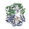 5xouC  5xowC  5xp8C 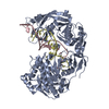 5xpaC 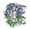 5xq2C 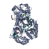 3dlhS S: Starting model for refinement C: citing same article ( |
|---|---|
| Similar structure data |


-Links
 PDBj
PDBj


- Assembly
| Deposited unit | 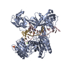
| ||||||||
|---|---|---|---|---|---|---|---|---|---|
| 1 |
| ||||||||
| Unit cell |
|
-Components
-Protein / DNA chain / RNA chain , 3 types, 3 molecules ACG
| #1: Protein | Mass: 76727.750 Da / Num. of mol.: 1 / Mutation: D546N Source method: isolated from a genetically manipulated source Source: (gene. exp.) Thermus thermophilus (strain HB27 / ATCC BAA-163 / DSM 7039) (bacteria)Strain: HB27 / ATCC BAA-163 / DSM 7039 / Gene: TT_P0026 / Plasmid: PET-SUMO / Production host: Escherichia coli (E. coli) / Strain (production host): Rosetta 2 (DE3)plyss / References: UniProt: Q746M7 |
|---|---|
| #2: DNA chain | Mass: 6588.266 Da / Num. of mol.: 1 / Source method: obtained synthetically / Source: (synth.) Thermus thermophilus (bacteria) |
| #3: RNA chain | Mass: 6271.791 Da / Num. of mol.: 1 / Source method: obtained synthetically / Source: (synth.) Thermus thermophilus (bacteria) |
-Non-polymers , 3 types, 26 molecules 


| #4: Chemical | Sulfate Mass: 96.063 Da / Num. of mol.: 2 / Source method: obtained synthetically / Formula: SO4 Mass: 96.063 Da / Num. of mol.: 2 / Source method: obtained synthetically / Formula: SO4#5: Chemical | ChemComp-MG / |  Mass: 24.305 Da / Num. of mol.: 1 / Source method: obtained synthetically / Formula: Mg Mass: 24.305 Da / Num. of mol.: 1 / Source method: obtained synthetically / Formula: Mg#6: Water | ChemComp-HOH / | WaterMass: 18.015 Da / Num. of mol.: 23 / Source method: isolated from a natural source / Formula: H2O |
|---|
-Experimental details
-Experiment
| Experiment | Method: X-RAY DIFFRACTION / Number of used crystals: 1 |
|---|
- Sample preparation
| Crystal | Density Matthews: 3.85 Å3/Da / Density % sol: 68.09 % |
|---|---|
| Crystal grow | Temperature: 308 K / Method: vapor diffusion, hanging drop / pH: 6.5 Details: 1.0 M (NH4)2SO4, 0.1 M KCL, 10 MM MGCL2, 50 MM BIS-TRIS PH 6.5 , VAPOR DIFFUSION, HANGING DROP, TEMPERATURE 308K PH range: 6.5 |
-Data collection
| Diffraction | Mean temperature: 100 K |
|---|---|
| Diffraction source | Source: SYNCHROTRON / Site: APS  / Beamline: 24-ID-C / Wavelength: 0.97926 Å / Beamline: 24-ID-C / Wavelength: 0.97926 Å |
| Detector | Type: MARMOSAIC 300 mm CCD / Detector: CCD / Date: Mar 15, 2009 |
| Radiation | Protocol: SINGLE WAVELENGTH / Monochromatic (M) / Laue (L): M / Scattering type: x-ray |
| Radiation wavelength | Wavelength: 0.97926 Å / Relative weight: 1 |
| Reflection | Resolution: 2.8→50 Å / Num. obs: 35223 / % possible obs: 99.5 % / Redundancy: 8.7 % / Biso Wilson estimate: 53.86 Å2 / Rmerge(I) obs: 0.086 / Net I/σ(I): 21.6 |
| Reflection shell | Resolution: 2.8→2.9 Å / Redundancy: 4.8 % / Rmerge(I) obs: 0.389 / Mean I/σ(I) obs: 2.2 / % possible all: 98.3 |
- Processing
| Software |
| ||||||||||||||||||||||||||||||||||||||||||||||||||||||||||||||||||||||||||||||||||||||||||||||||||
|---|---|---|---|---|---|---|---|---|---|---|---|---|---|---|---|---|---|---|---|---|---|---|---|---|---|---|---|---|---|---|---|---|---|---|---|---|---|---|---|---|---|---|---|---|---|---|---|---|---|---|---|---|---|---|---|---|---|---|---|---|---|---|---|---|---|---|---|---|---|---|---|---|---|---|---|---|---|---|---|---|---|---|---|---|---|---|---|---|---|---|---|---|---|---|---|---|---|---|---|
| Refinement | Method to determine structure: MOLECULAR REPLACEMENT Starting model: 3DLH Resolution: 2.8→49.08 Å / SU ML: 0.4 / Cross valid method: FREE R-VALUE / σ(F): 0.16
| ||||||||||||||||||||||||||||||||||||||||||||||||||||||||||||||||||||||||||||||||||||||||||||||||||
| Solvent computation | Bsol: 43.9 Å2 / ksol: 0.34 e/Å3 | ||||||||||||||||||||||||||||||||||||||||||||||||||||||||||||||||||||||||||||||||||||||||||||||||||
| Displacement parameters | Biso mean: 57.19 Å2
| ||||||||||||||||||||||||||||||||||||||||||||||||||||||||||||||||||||||||||||||||||||||||||||||||||
| Refinement step | Cycle: LAST / Resolution: 2.8→49.08 Å
| ||||||||||||||||||||||||||||||||||||||||||||||||||||||||||||||||||||||||||||||||||||||||||||||||||
| Refine LS restraints |
| ||||||||||||||||||||||||||||||||||||||||||||||||||||||||||||||||||||||||||||||||||||||||||||||||||
| LS refinement shell |
|
