[English] 日本語
Yorodumi
- PDB-4r1m: Crystal structure of a Putative Acyl-CoA ligase (BT_0428) from Ba... -

+
Open data


ID or keywords:

Loading...

-
Basic information

Entry
Database: PDB / ID: 4r1m
TitleCrystal structure of a Putative Acyl-CoA ligase (BT_0428) from Bacteroides thetaiotaomicron VPI-5482 at 2.48 A resolution
ComponentsPhenylacetate-coenzyme A ligase
KeywordsLIGASE / Acetyl-CoA synthetase-like / Structural Genomics / Joint Center for Structural Genomics / JCSG / Protein Structure Initiative / PSI-BIOLOGY
Function / homology
Function and homology information


phenylacetate-CoA ligase / phenylacetate-CoA ligase activity / phenylacetate catabolic process / nucleotide binding / metal ion binding
Similarity search - Function
Phenylacetate-CoA ligase / AMP-dependent ligase, C-terminal / AMP-binding enzyme C-terminal domain / ANL, C-terminal domain / ANL, N-terminal domain / ANL, N-terminal domain / GMP Synthetase; Chain A, domain 3 / AMP-dependent synthetase/ligase / AMP-binding enzyme, C-terminal domain superfamily / AMP-binding enzyme ...Phenylacetate-CoA ligase / AMP-dependent ligase, C-terminal / AMP-binding enzyme C-terminal domain / ANL, C-terminal domain / ANL, N-terminal domain / ANL, N-terminal domain / GMP Synthetase; Chain A, domain 3 / AMP-dependent synthetase/ligase / AMP-binding enzyme, C-terminal domain superfamily / AMP-binding enzyme / Rossmann fold / 2-Layer Sandwich / 3-Layer(aba) Sandwich / Alpha Beta
Similarity search - Domain/homology
ADENOSINE MONOPHOSPHATE / Phenylacetate-coenzyme A ligase
Similarity search - Component
Biological speciesBacteroides thetaiotaomicron (bacteria)
MethodX-RAY DIFFRACTION / SYNCHROTRON / SAD / Resolution: 2.48 Å
AuthorsJoint Center for Structural Genomics (JCSG)
CitationJournal: To be published
Title: Crystal structure of a Hypothetical Acyl-CoA ligase (BT_0428) from Bacteroides thetaiotaomicron VPI-5482 at 2.48 A resolution
Authors: Joint Center for Structural Genomics (JCSG)
History
DepositionAug 6, 2014Deposition site: RCSB / Processing site: RCSB
Revision 1.0Aug 27, 2014Provider: repository / Type: Initial release
Revision 1.1Dec 24, 2014Group: Structure summary
Revision 1.2Nov 22, 2017Group: Refinement description / Category: software
Revision 1.3Jan 24, 2018Group: Database references / Category: citation_author / Item: _citation_author.name
Revision 1.4Feb 1, 2023Group: Database references / Derived calculations
Category: database_2 / pdbx_struct_conn_angle ...database_2 / pdbx_struct_conn_angle / struct_conn / struct_ref_seq_dif / struct_site
Item: _database_2.pdbx_DOI / _database_2.pdbx_database_accession ..._database_2.pdbx_DOI / _database_2.pdbx_database_accession / _pdbx_struct_conn_angle.ptnr1_auth_asym_id / _pdbx_struct_conn_angle.ptnr1_auth_comp_id / _pdbx_struct_conn_angle.ptnr1_auth_seq_id / _pdbx_struct_conn_angle.ptnr1_label_asym_id / _pdbx_struct_conn_angle.ptnr1_label_atom_id / _pdbx_struct_conn_angle.ptnr1_label_comp_id / _pdbx_struct_conn_angle.ptnr1_label_seq_id / _pdbx_struct_conn_angle.ptnr2_auth_asym_id / _pdbx_struct_conn_angle.ptnr2_auth_comp_id / _pdbx_struct_conn_angle.ptnr2_auth_seq_id / _pdbx_struct_conn_angle.ptnr2_label_asym_id / _pdbx_struct_conn_angle.ptnr2_label_atom_id / _pdbx_struct_conn_angle.ptnr2_label_comp_id / _pdbx_struct_conn_angle.ptnr3_auth_asym_id / _pdbx_struct_conn_angle.ptnr3_auth_comp_id / _pdbx_struct_conn_angle.ptnr3_auth_seq_id / _pdbx_struct_conn_angle.ptnr3_label_asym_id / _pdbx_struct_conn_angle.ptnr3_label_atom_id / _pdbx_struct_conn_angle.ptnr3_label_comp_id / _pdbx_struct_conn_angle.ptnr3_label_seq_id / _pdbx_struct_conn_angle.value / _struct_conn.pdbx_dist_value / _struct_conn.pdbx_leaving_atom_flag / _struct_conn.ptnr1_auth_asym_id / _struct_conn.ptnr1_auth_comp_id / _struct_conn.ptnr1_auth_seq_id / _struct_conn.ptnr1_label_asym_id / _struct_conn.ptnr1_label_atom_id / _struct_conn.ptnr1_label_comp_id / _struct_conn.ptnr1_label_seq_id / _struct_conn.ptnr2_auth_asym_id / _struct_conn.ptnr2_auth_comp_id / _struct_conn.ptnr2_auth_seq_id / _struct_conn.ptnr2_label_asym_id / _struct_conn.ptnr2_label_atom_id / _struct_conn.ptnr2_label_comp_id / _struct_conn.ptnr2_label_seq_id / _struct_ref_seq_dif.details / _struct_site.pdbx_auth_asym_id / _struct_site.pdbx_auth_comp_id / _struct_site.pdbx_auth_seq_id
Revision 1.5Nov 29, 2023Group: Data collection / Database references
Category: chem_comp_atom / chem_comp_bond / pdbx_database_related
Item: _pdbx_database_related.db_name

-
Structure visualization

Structure viewerMolecule:
MolmilJmol/JSmol

Downloads & links

-
Assembly

Deposited unit
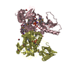
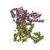
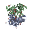
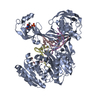
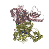
A: Phenylacetate-coenzyme A ligase
B: Phenylacetate-coenzyme A ligase
C: Phenylacetate-coenzyme A ligase
D: Phenylacetate-coenzyme A ligase
hetero molecules


Theoretical massNumber of molelcules
Total (without water)203,21332
Polymers200,8164
Non-polymers2,39728
Water5,062281
1
A: Phenylacetate-coenzyme A ligase
B: Phenylacetate-coenzyme A ligase
hetero molecules


Theoretical massNumber of molelcules
Total (without water)101,90018
Polymers100,4082
Non-polymers1,49216
Water362
TypeNameSymmetry operationNumber
identity operation1_555x,y,z1
Buried area7320 Å2
ΔGint-20 kcal/mol
Surface area34480 Å2
MethodPISA
2
C: Phenylacetate-coenzyme A ligase
D: Phenylacetate-coenzyme A ligase
hetero molecules


Theoretical massNumber of molelcules
Total (without water)101,31314
Polymers100,4082
Non-polymers90512
Water362
TypeNameSymmetry operationNumber
identity operation1_555x,y,z1
Buried area5360 Å2
ΔGint-40 kcal/mol
Surface area34910 Å2
MethodPISA
Unit cell
Length a, b, c (Å)127.060, 211.870, 72.250
Angle α, β, γ (deg.)90.000, 90.000, 90.000
Int Tables number18
Space group name H-MP21212

-
Components

-
Protein , 1 types, 4 molecules ABCD

#1: Protein
Phenylacetate-coenzyme A ligase


Mass: 50203.996 Da / Num. of mol.: 4
Source method: isolated from a genetically manipulated source
Source: (gene. exp.) Bacteroides thetaiotaomicron (bacteria)
Strain: VPI-5482 / Gene: BT_0428 / Plasmid: SpeedET / Production host: Escherichia Coli (E. coli) / Strain (production host): HK100 / References: UniProt: Q8AAN6

-
Non-polymers , 6 types, 309 molecules

#2: Chemical
ChemComp-ZN / ZINC ION


Mass: 65.409 Da / Num. of mol.: 4 / Source method: obtained synthetically / Formula: Zn
#3: Chemical ChemComp-AMP / ADENOSINE MONOPHOSPHATE / Adenosine monophosphate


Mass: 347.221 Da / Num. of mol.: 3 / Source method: obtained synthetically / Formula: C10H14N5O7P / Comment: AMP*YM
#4: Chemical
ChemComp-NA / SODIUM ION


Mass: 22.990 Da / Num. of mol.: 4 / Source method: obtained synthetically / Formula: Na
#5: Chemical
ChemComp-EDO / 1,2-ETHANEDIOL / ETHYLENE GLYCOL / Ethylene glycol


Mass: 62.068 Da / Num. of mol.: 15 / Source method: obtained synthetically / Formula: C2H6O2
#6: Chemical ChemComp-CL / CHLORIDE ION / Chloride


Mass: 35.453 Da / Num. of mol.: 2 / Source method: obtained synthetically / Formula: Cl
#7: Water ChemComp-HOH / water / Water


Mass: 18.015 Da / Num. of mol.: 281 / Source method: isolated from a natural source / Formula: H2O

-
Details

Sequence detailsTHE CONSTRUCT (RESIDUES 1-435) WAS EXPRESSED WITH A PURIFICATION TAG MGSDKIHHHHHHENLYFQG. THE TAG ...THE CONSTRUCT (RESIDUES 1-435) WAS EXPRESSED WITH A PURIFICATION TAG MGSDKIHHHHHHENLYFQG. THE TAG WAS REMOVED WITH TEV PROTEASE LEAVING ONLY A GLYCINE (0) FOLLOWED BY THE TARGET SEQUENCE.

-
Experimental details

-
Experiment

ExperimentMethod: X-RAY DIFFRACTION / Number of used crystals: 1

-
Sample preparation

CrystalDensity Matthews: 2.42 Å3/Da / Density % sol: 49.2 %
Crystal growTemperature: 293 K / Method: vapor diffusion, sitting drop / pH: 7.9
Details: 16.0% polyethylene glycol 3000, 0.25M sodium chloride, 0.1M HEPES pH 7.9, 5mM Phenylacetate, 5mM MgCl2, 3mM Coenzyme A, NANODROP, VAPOR DIFFUSION, SITTING DROP, temperature 293K

-
Data collection

DiffractionMean temperature: 100 K
Diffraction sourceSource: SYNCHROTRON / Site: SSRL / Beamline: BL11-1 / Wavelength: 0.97954
DetectorType: MARMOSAIC 325 mm CCD / Detector: CCD / Date: Nov 23, 2010
Details: Flat mirror (vertical focusing); single crystal Si(111) bent monochromator (horizontal focusing)
RadiationMonochromator: single crystal Si(111) bent / Protocol: SINGLE WAVELENGTH / Monochromatic (M) / Laue (L): M / Scattering type: x-ray
Radiation wavelengthWavelength: 0.97954 Å / Relative weight: 1
ReflectionResolution: 2.48→48.889 Å / Num. obs: 69731 / % possible obs: 99.5 % / Observed criterion σ(I): -3 / Biso Wilson estimate: 59.47 Å2 / Rmerge(I) obs: 0.077 / Net I/σ(I): 12.33
Reflection shell

Diffraction-ID: 1

Resolution (Å)Highest resolution (Å)Rmerge(I) obsMean I/σ(I) obsNum. measured obsNum. unique obs% possible all
2.48-2.570.7242.234167698099.2
2.57-2.670.5412.9335496714100
2.67-2.790.4023.834063682799.9
2.79-2.940.2815.335205704099.9
2.94-3.120.208734041684399.9
3.12-3.360.12110.934419693299.9
3.36-3.70.0751634661704499.6
3.7-4.230.05121.633989697899.4
4.23-5.310.04625.233778699399.1
5.310.04827.133878738097.9

-
Phasing

PhasingMethod: SAD

-
Processing

Software
NameVersionClassificationNB
MolProbity3beta29model building
PDB_EXTRACT3.1data extraction
SHARPphasing
XSCALEJuly 4, 2012 BUILT=20130617data scaling
BUSTER-TNT2.10.0refinement
XDSdata reduction
BUSTER2.10.0refinement
RefinementMethod to determine structure: SAD / Resolution: 2.48→48.889 Å / Cor.coef. Fo:Fc: 0.9494 / Cor.coef. Fo:Fc free: 0.939 / Occupancy max: 1 / Occupancy min: 0.5 / Cross valid method: THROUGHOUT / σ(F): 0
Details: 1.A MET-INHIBITION PROTOCOL WAS USED FOR SELENOMETHIONINE INCORPORATION DURING PROTEIN EXPRESSION. THE OCCUPANCY OF THE SE ATOMS IN THE MSE RESIDUES WAS REDUCED TO 0.75 FOR THE REDUCED ...Details: 1.A MET-INHIBITION PROTOCOL WAS USED FOR SELENOMETHIONINE INCORPORATION DURING PROTEIN EXPRESSION. THE OCCUPANCY OF THE SE ATOMS IN THE MSE RESIDUES WAS REDUCED TO 0.75 FOR THE REDUCED SCATTERING POWER DUE TO PARTIAL S-MET INCORPORATION. 2.PROTEIN ATOM RECORD CONTAINS SUM OF TLS AND RESIDUAL B FACTORS. ANISOU RECORD CONTAINS SUM OF TLS AND RESIDUAL U FACTORS. 3. NCS RESTRAINTS WERE APPLIED USING BUSTER'S LSSR RESTRAINT REPRESENTATION. 4. THE MODELING OF ZINC IS IS SUPPORTED BY ANOMALOUS DIFFERENCE MAPS AND SIMILAR CRYSTALS OF THE SAME PROTEIN. 5. SODIUM (NA) AND CHLORIDE (CL) FROM THE CRYSTALLIZATION WERE MODELED INTO THE STRUCTURE. 1,2-ETHANEDIOL (EDO) USED AS A CRYOPROTECTANT WAS MODELED INTO THE STRUCTURE. 6. ADENOSINE-5'-MONOSPHATE (AMP),MOST LIKELY BOUND TO THE PROTEIN DURING EXPRESSION WAS MODELED INTO THE ACTIVE SITE ON SUBUNITS A,B, AND D IN THE ASYMMETRIC UNIT. HOWEVER,ELECTRON DENSITY FOR AMP WAS ABSENT ON SUBUNIT C; THEREFORE AMO COULD NOT BE RELIABLY MODELED INTO THE ACTIVE SITE OF THIS SUBUNIT. 7. ASN 243 ON THE A,B,C, AND D-CHAINS ARE RAMACHANDRAN OUTLIERS IN MOLPROBITY EVEN THOUGH THEIR POSITIONINGS ARE SUPPORTED BY ELECTRON DENSITY. LYS 84 ON THE C-CHAIN IS A RAMACHANDRAN OUTLIER EVEN THOUGH ITS POSITIONING IS SUPPORTED BY ELECTRON DENSITY. SER 382 AND ASN 367 ON THE B CHAIN, AND VAL 366 ON THE D CHAIN ARE IN POOR REGIONS OF ELECTRON DENSITY AND ARE FLAGGED AS RAMACHANDRAN OUTLIERS IN MOLPROBITY.
RfactorNum. reflection% reflectionSelection details
Rfree0.2001 3483 5 %RANDOM
Rwork0.1748 ---
obs0.1761 69658 99.47 %-
Displacement parametersBiso max: 161.42 Å2 / Biso mean: 67.5728 Å2 / Biso min: 30.91 Å2
Baniso -1Baniso -2Baniso -3
1--12.8647 Å20 Å20 Å2
2--7.9879 Å20 Å2
3---4.8769 Å2
Refine analyzeLuzzati coordinate error obs: 0.355 Å
Refinement stepCycle: LAST / Resolution: 2.48→48.889 Å
ProteinNucleic acidLigandSolventTotal
Num. atoms13380 0 139 281 13800
Refine LS restraints
Refine-IDTypeNumberRestraint functionWeightDev ideal
X-RAY DIFFRACTIONt_dihedral_angle_d6538SINUSOIDAL8
X-RAY DIFFRACTIONt_trig_c_planes355HARMONIC2
X-RAY DIFFRACTIONt_gen_planes2043HARMONIC5
X-RAY DIFFRACTIONt_it13802HARMONIC20
X-RAY DIFFRACTIONt_nbd0SEMIHARMONIC5
X-RAY DIFFRACTIONt_improper_torsion
X-RAY DIFFRACTIONt_pseud_angle
X-RAY DIFFRACTIONt_chiral_improper_torsion1857SEMIHARMONIC6
X-RAY DIFFRACTIONt_sum_occupancies
X-RAY DIFFRACTIONt_utility_distance
X-RAY DIFFRACTIONt_utility_angle
X-RAY DIFFRACTIONt_utility_torsion
X-RAY DIFFRACTIONt_ideal_dist_contact15468SEMIHARMONIC4
X-RAY DIFFRACTIONt_bond_d13802HARMONIC20.01
X-RAY DIFFRACTIONt_angle_deg18662HARMONIC21.09
X-RAY DIFFRACTIONt_omega_torsion3.22
X-RAY DIFFRACTIONt_other_torsion2.21
LS refinement shellResolution: 2.48→2.54 Å / Total num. of bins used: 20
RfactorNum. reflection% reflection
Rfree0.2602 208 4.1 %
Rwork0.2132 4862 -
all0.2151 5070 -
obs--99.47 %
Refinement TLS params.

Method: refined / Refine-ID: X-RAY DIFFRACTION

IDL112)L122)L132)L222)L232)L332)S11 (Å °)S12 (Å °)S13 (Å °)S21 (Å °)S22 (Å °)S23 (Å °)S31 (Å °)S32 (Å °)S33 (Å °)T112)T122)T132)T222)T232)T332)Origin x (Å)Origin y (Å)Origin z (Å)
11.03910.813-0.17752.2256-0.74372.292-0.03540.09860.0845-0.10880.04150.0982-0.0115-0.1733-0.0061-0.0523-0.02360.0283-0.11090.0134-0.1446-0.29461.358727.6456
24.3096-2.91040.38995.0538-1.44732.72740.07640.2496-0.34520.18-0.2236-0.54420.020.16030.1472-0.2137-0.04830.0072-0.18270.1520.181926.241747.709828.0211
31.45381.2709-0.09372.8786-0.69131.63810.0335-0.08670.08710.5442-0.03980.1555-0.2194-0.10250.00620.02110.00150.0416-0.17430.0108-0.1665-4.366248.494259.5207
48.188-2.91042.16884.863-0.5351.6987-0.0081-0.077-0.536-0.084-0.187-0.24550.16310.09630.1951-0.09720.0354-0.1071-0.3040.1520.106819.11532.804967.4045
52.14410.20990.21231.36460.55323.00910.0975-0.4008-0.06730.3483-0.083-0.16440.54420.0679-0.01460.0078-0.0189-0.0094-0.1799-0.0148-0.248638.5132-11.207575.2928
63.4651-2.28130.04718.246-1.88833.5552-0.0523-0.20790.54420.27020.12720.0692-0.2756-0.0195-0.0749-0.2108-0.0219-0.0336-0.1379-0.0737-0.021141.36717.965874.7899
72.48680.44030.61721.46220.09621.44430.04870.23350.1119-0.254-0.0190.03650.1934-0.0793-0.02960.05250.0189-0.0003-0.1539-0.0089-0.181326.1425-7.494843.2463
81.6079-1.2769-1.04378.3155-1.81162.02570.00150.23740.5294-0.1637-0.34940.1496-0.2346-0.17720.3479-0.04720.1079-0.152-0.27840.1520.04625.650921.602535.3756
Refinement TLS group
IDRefine-IDRefine TLS-IDSelection detailsAuth asym-IDAuth seq-ID
1X-RAY DIFFRACTION1{A|0 - 325}A0 - 325
2X-RAY DIFFRACTION2{A|326 - 434}A326 - 434
3X-RAY DIFFRACTION3{B|3 - 327}B3 - 327
4X-RAY DIFFRACTION4{B|328 - 432}B328 - 432
5X-RAY DIFFRACTION5{C|0 - 323}C0 - 323
6X-RAY DIFFRACTION6{C|324 - 432}C324 - 432
7X-RAY DIFFRACTION7{D|2 - 333}D2 - 333
8X-RAY DIFFRACTION8{D|334 - 434}D334 - 434

+
About Yorodumi

-
News

-
Feb 9, 2022. New format data for meta-information of EMDB entries

New format data for meta-information of EMDB entries

  • Version 3 of the EMDB header file is now the official format.
  • The previous official version 1.9 will be removed from the archive.

Related info.:EMDB header

External links:wwPDB to switch to version 3 of the EMDB data model

-
Aug 12, 2020. Covid-19 info

Covid-19 info

URL: https://pdbj.org/emnavi/covid19.php

New page: Covid-19 featured information page in EM Navigator.

Related info.:Covid-19 info / Mar 5, 2020. Novel coronavirus structure data

+
Mar 5, 2020. Novel coronavirus structure data

Novel coronavirus structure data

Related info.:Yorodumi Speices / Aug 12, 2020. Covid-19 info

External links:COVID-19 featured content - PDBj / Molecule of the Month (242):Coronavirus Proteases

+
Jan 31, 2019. EMDB accession codes are about to change! (news from PDBe EMDB page)

EMDB accession codes are about to change! (news from PDBe EMDB page)

  • The allocation of 4 digits for EMDB accession codes will soon come to an end. Whilst these codes will remain in use, new EMDB accession codes will include an additional digit and will expand incrementally as the available range of codes is exhausted. The current 4-digit format prefixed with “EMD-” (i.e. EMD-XXXX) will advance to a 5-digit format (i.e. EMD-XXXXX), and so on. It is currently estimated that the 4-digit codes will be depleted around Spring 2019, at which point the 5-digit format will come into force.
  • The EM Navigator/Yorodumi systems omit the EMD- prefix.

Related info.:Q: What is EMD? / ID/Accession-code notation in Yorodumi/EM Navigator

External links:EMDB Accession Codes are Changing Soon! / Contact to PDBj

+
Jul 12, 2017. Major update of PDB

Major update of PDB

  • wwPDB released updated PDB data conforming to the new PDBx/mmCIF dictionary.
  • This is a major update changing the version number from 4 to 5, and with Remediation, in which all the entries are updated.
  • In this update, many items about electron microscopy experimental information are reorganized (e.g. em_software).
  • Now, EM Navigator and Yorodumi are based on the updated data.

External links:wwPDB Remediation / Enriched Model Files Conforming to OneDep Data Standards Now Available in the PDB FTP Archive

-
Yorodumi

Thousand views of thousand structures

  • Yorodumi is a browser for structure data from EMDB, PDB, SASBDB, etc.
  • This page is also the successor to EM Navigator detail page, and also detail information page/front-end page for Omokage search.
  • The word "yorodu" (or yorozu) is an old Japanese word meaning "ten thousand". "mi" (miru) is to see.

Related info.:EMDB / PDB / SASBDB / Comparison of 3 databanks / Yorodumi Search / Aug 31, 2016. New EM Navigator & Yorodumi / Yorodumi Papers / Jmol/JSmol / Function and homology information / Changes in new EM Navigator and Yorodumi

Read more