 Movie
Movie Controller

[English] 日本語
 Yorodumi
- PDB-5xmc: Crystal structure of the auto-inhibited Nedd4 family E3 ligase Itch -
+ Open data

- Basic information
| Entry | Database: PDB / ID: 5xmc | ||||||
|---|---|---|---|---|---|---|---|
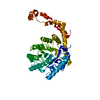
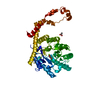
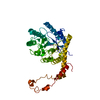
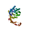
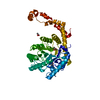
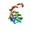
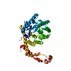
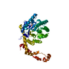
| Title | Crystal structure of the auto-inhibited Nedd4 family E3 ligase Itch | ||||||
 Components Components | E3 ubiquitin-protein ligase Itchy | ||||||
 Keywords Keywords |  LIGASE / Auto-inhibited Itch LIGASE / Auto-inhibited Itch | ||||||
| Function / homology |  Function and homology information Function and homology informationregulation of protein deubiquitination / Downregulation of ERBB4 signaling / negative regulation of cytoplasmic pattern recognition receptor signaling pathway / Activated NOTCH1 Transmits Signal to the Nucleus / Regulation of necroptotic cell death / negative regulation of defense response to virus / Degradation of GLI1 by the proteasome / protein K29-linked ubiquitination / Hedgehog 'on' state / T cell anergy ...regulation of protein deubiquitination / Downregulation of ERBB4 signaling / negative regulation of cytoplasmic pattern recognition receptor signaling pathway / Activated NOTCH1 Transmits Signal to the Nucleus / Regulation of necroptotic cell death / negative regulation of defense response to virus / Degradation of GLI1 by the proteasome / protein K29-linked ubiquitination / Hedgehog 'on' state / T cell anergy / positive regulation of T cell anergy / RUNX1 regulates transcription of genes involved in differentiation of HSCs / CXCR chemokine receptor binding / CD4-positive, alpha-beta T cell proliferation / NOD1/2 Signaling Pathway / Antigen processing: Ubiquitination & Proteasome degradation / negative regulation of CD4-positive, alpha-beta T cell proliferation / HECT-type E3 ubiquitin transferase / negative regulation of JNK cascade / arrestin family protein binding / positive regulation of receptor catabolic process / negative regulation of NF-kappaB transcription factor activity / protein monoubiquitination / ubiquitin-like protein ligase binding / protein K63-linked ubiquitination / ligase activity / protein K48-linked ubiquitination / protein autoubiquitination / ubiquitin ligase complex / ribonucleoprotein complex binding / protein catabolic process / receptor internalization / protein polyubiquitination / ubiquitin-protein transferase activity / positive regulation of protein catabolic process / ubiquitin protein ligase activity / cell cortex / ubiquitin-dependent protein catabolic process / cytoplasmic vesicle / early endosome membrane / proteasome-mediated ubiquitin-dependent protein catabolic process / defense response to virus / protein ubiquitination / intracellular membrane-bounded organelle / innate immune response / apoptotic process / negative regulation of apoptotic process / protein-containing complex / nucleoplasm / membrane / nucleus / plasma membrane / cytosol / cytoplasmSimilarity search - Function | ||||||
| Biological species |  Mus musculus (house mouse) Mus musculus (house mouse) | ||||||
| Method | X-RAY DIFFRACTION / SYNCHROTRON / MOLECULAR REPLACEMENT / molecular replacement / Resolution: 2.6 Å | ||||||
| Model details | Allosteric auto-inhibition and activation of Nedd4 family E3 ligase Itch | ||||||
 Authors Authors | Shan, Z. / Wen, W. | ||||||
 Citation Citation | Journal: EMBO Rep. / Year: 2017 Title: Allosteric auto-inhibition and activation of the Nedd4 family E3 ligase Itch Authors: Zhu, K. / Shan, Z. / Chen, X. / Cai, Y. / Cui, L. / Yao, W. / Wang, Z. / Shi, P. / Tian, C. / Lou, J. / Xie, Y. / Wen, W. | ||||||
| History |
|
- Structure visualization
| Structure viewer | Molecule: MolmilJmol/JSmol |
|---|
- Downloads & links
-Download
| PDBx/mmCIF format | 5xmc.cif.gz | 189.7 KB | Display | PDBx/mmCIF format |
|---|---|---|---|---|
| PDB format | pdb5xmc.ent.gz | 144.5 KB | Display | PDB format |
| PDBx/mmJSON format | 5xmc.json.gz | Tree view | PDBx/mmJSON format | |
| Others |  Other downloads Other downloads |
-Validation report
| Arichive directory | https://data.pdbj.org/pub/pdb/validation_reports/xm/5xmcftp://data.pdbj.org/pub/pdb/validation_reports/xm/5xmc | HTTPS FTP |
|---|
-Related structure data
| Related structure data |  3tugS S: Starting model for refinement |
|---|---|
| Similar structure data |


-Links
 PDBj
PDBj


- Assembly
| Deposited unit | 
| ||||||||
|---|---|---|---|---|---|---|---|---|---|
| 1 |
| ||||||||
| Unit cell |
|
-Components
| #1: Protein | Mass: 83412.203 Da / Num. of mol.: 1 / Fragment: Nedd4 family E3 ligase Itch, UNP residues 143-864 Source method: isolated from a genetically manipulated source Source: (gene. exp.) Mus musculus (house mouse) / Gene: Itch / Production host:  Escherichia coli BL21 (bacteria) / Strain (production host): BL21 Escherichia coli BL21 (bacteria) / Strain (production host): BL21References: UniProt: Q8C863, HECT-type E3 ubiquitin transferase |
|---|---|
| #2: Water | ChemComp-HOH / Water Mass: 18.015 Da / Num. of mol.: 46 / Source method: isolated from a natural source / Formula: H2O Mass: 18.015 Da / Num. of mol.: 46 / Source method: isolated from a natural source / Formula: H2O |
-Experimental details
-Experiment
| Experiment | Method: X-RAY DIFFRACTION / Number of used crystals: 1 |
|---|
- Sample preparation
| Crystal | Density Matthews: 2.1 Å3/Da / Density % sol: 41.36 % |
|---|---|
| Crystal grow | Temperature: 289 K / Method: evaporation / pH: 5.5 Details: 50mM citric acid, 50mM Bis-Tris propone, pH 5.5, 16% PEG 3350 |
-Data collection
| Diffraction | Mean temperature: 100 K | ||||||||||||||||||||||||||||||||||||||||||||||||||||||||||||||||||||||||||||||||||||||||||||||||||||||||||||||||||||||||||||||||||||||||||||||||||||||||||||||||||||||||
|---|---|---|---|---|---|---|---|---|---|---|---|---|---|---|---|---|---|---|---|---|---|---|---|---|---|---|---|---|---|---|---|---|---|---|---|---|---|---|---|---|---|---|---|---|---|---|---|---|---|---|---|---|---|---|---|---|---|---|---|---|---|---|---|---|---|---|---|---|---|---|---|---|---|---|---|---|---|---|---|---|---|---|---|---|---|---|---|---|---|---|---|---|---|---|---|---|---|---|---|---|---|---|---|---|---|---|---|---|---|---|---|---|---|---|---|---|---|---|---|---|---|---|---|---|---|---|---|---|---|---|---|---|---|---|---|---|---|---|---|---|---|---|---|---|---|---|---|---|---|---|---|---|---|---|---|---|---|---|---|---|---|---|---|---|---|---|---|---|---|
| Diffraction source | Source: SYNCHROTRON / Site: SSRF  / Beamline: BL18U1 / Wavelength: 0.975 Å / Beamline: BL18U1 / Wavelength: 0.975 Å | ||||||||||||||||||||||||||||||||||||||||||||||||||||||||||||||||||||||||||||||||||||||||||||||||||||||||||||||||||||||||||||||||||||||||||||||||||||||||||||||||||||||||
| Detector | Type: DECTRIS PILATUS3 6M / Detector: PIXEL / Date: Mar 23, 2015 | ||||||||||||||||||||||||||||||||||||||||||||||||||||||||||||||||||||||||||||||||||||||||||||||||||||||||||||||||||||||||||||||||||||||||||||||||||||||||||||||||||||||||
| Radiation | Monochromator: double crystal / Protocol: SINGLE WAVELENGTH / Monochromatic (M) / Laue (L): M / Scattering type: x-ray | ||||||||||||||||||||||||||||||||||||||||||||||||||||||||||||||||||||||||||||||||||||||||||||||||||||||||||||||||||||||||||||||||||||||||||||||||||||||||||||||||||||||||
| Radiation wavelength | Wavelength: 0.975 Å / Relative weight: 1 | ||||||||||||||||||||||||||||||||||||||||||||||||||||||||||||||||||||||||||||||||||||||||||||||||||||||||||||||||||||||||||||||||||||||||||||||||||||||||||||||||||||||||
| Reflection | Resolution: 2.6→50 Å / Num. obs: 22580 / % possible obs: 99.7 % / Redundancy: 4.7 % / Biso Wilson estimate: 38.2 Å2 / Rmerge(I) obs: 0.071 / Rpim(I) all: 0.036 / Rrim(I) all: 0.08 / Χ2: 0.895 / Net I/σ(I): 6.7 | ||||||||||||||||||||||||||||||||||||||||||||||||||||||||||||||||||||||||||||||||||||||||||||||||||||||||||||||||||||||||||||||||||||||||||||||||||||||||||||||||||||||||
| Reflection shell | Diffraction-ID: 1
|
-Phasing
| Phasing | Method: molecular replacement |
|---|
- Processing
| Software |
| |||||||||||||||||||||||||||||||||||||||||||||||||||||||||||||||||||||||||||||||||||||||||||||||||||||||||||||||||||||||||||||
|---|---|---|---|---|---|---|---|---|---|---|---|---|---|---|---|---|---|---|---|---|---|---|---|---|---|---|---|---|---|---|---|---|---|---|---|---|---|---|---|---|---|---|---|---|---|---|---|---|---|---|---|---|---|---|---|---|---|---|---|---|---|---|---|---|---|---|---|---|---|---|---|---|---|---|---|---|---|---|---|---|---|---|---|---|---|---|---|---|---|---|---|---|---|---|---|---|---|---|---|---|---|---|---|---|---|---|---|---|---|---|---|---|---|---|---|---|---|---|---|---|---|---|---|---|---|---|
| Refinement | Method to determine structure: MOLECULAR REPLACEMENT Starting model: 3TUG Resolution: 2.6→47.593 Å / SU ML: 0.34 / Cross valid method: FREE R-VALUE / σ(F): 1.36 / Phase error: 25.95
| |||||||||||||||||||||||||||||||||||||||||||||||||||||||||||||||||||||||||||||||||||||||||||||||||||||||||||||||||||||||||||||
| Solvent computation | Shrinkage radii: 0.9 Å / VDW probe radii: 1.11 Å | |||||||||||||||||||||||||||||||||||||||||||||||||||||||||||||||||||||||||||||||||||||||||||||||||||||||||||||||||||||||||||||
| Displacement parameters | Biso max: 114.9 Å2 / Biso mean: 46.12 Å2 / Biso min: 12.56 Å2 | |||||||||||||||||||||||||||||||||||||||||||||||||||||||||||||||||||||||||||||||||||||||||||||||||||||||||||||||||||||||||||||
| Refinement step | Cycle: final / Resolution: 2.6→47.593 Å
| |||||||||||||||||||||||||||||||||||||||||||||||||||||||||||||||||||||||||||||||||||||||||||||||||||||||||||||||||||||||||||||
| Refine LS restraints |
| |||||||||||||||||||||||||||||||||||||||||||||||||||||||||||||||||||||||||||||||||||||||||||||||||||||||||||||||||||||||||||||
| LS refinement shell | Refine-ID: X-RAY DIFFRACTION / Rfactor Rfree error: 0 / Total num. of bins used: 8
| |||||||||||||||||||||||||||||||||||||||||||||||||||||||||||||||||||||||||||||||||||||||||||||||||||||||||||||||||||||||||||||
| Refinement TLS params. | Method: refined / Refine-ID: X-RAY DIFFRACTION
| |||||||||||||||||||||||||||||||||||||||||||||||||||||||||||||||||||||||||||||||||||||||||||||||||||||||||||||||||||||||||||||
| Refinement TLS group |
|
