 Movie
Movie Controller


+ Open data

- Basic information
| Entry | Database: PDB / ID: 5wwc | ||||||
|---|---|---|---|---|---|---|---|
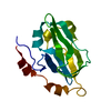
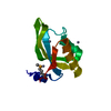
| Title | The crystal structure of Cren7 mutant L28M in complex with dsDNA | ||||||
 Components Components |
| ||||||
 Keywords Keywords | DNA BINDING PROTEIN/DNA /  BETA-SHEET / DNA BINDING / DNA BINDING PROTEIN-DNA COMPLEX / Crenarchaeal chromatin protein BETA-SHEET / DNA BINDING / DNA BINDING PROTEIN-DNA COMPLEX / Crenarchaeal chromatin protein | ||||||
| Function / homology |  Function and homology information Function and homology information | ||||||
| Biological species |   Sulfolobus solfataricus (archaea) Sulfolobus solfataricus (archaea)synthetic construct (others) | ||||||
| Method | X-RAY DIFFRACTION / SYNCHROTRON / MOLECULAR REPLACEMENT / Resolution: 1.9 Å | ||||||
 Authors Authors | Zhang, Z.F. / Zhao, M.H. / Wang, L. / Chen, Y.Y. / Dong, Y.H. / Gong, Y. / Huang, L. | ||||||
 Citation Citation | Journal: Biochem. J. / Year: 2017 Title: Roles of Leu28 side chain intercalation in the interaction between Cren7 and DNA Authors: Zhang, Z. / Zhao, M. / Wang, L. / Chen, Y. / Dong, Y. / Gong, Y. / Huang, L. | ||||||
| History |
|
- Structure visualization
| Structure viewer | Molecule: MolmilJmol/JSmol |
|---|
- Downloads & links
-Download
| PDBx/mmCIF format | 5wwc.cif.gz | 57.2 KB | Display | PDBx/mmCIF format |
|---|---|---|---|---|
| PDB format | pdb5wwc.ent.gz | 37.3 KB | Display | PDB format |
| PDBx/mmJSON format | 5wwc.json.gz | Tree view | PDBx/mmJSON format | |
| Others |  Other downloads Other downloads |
-Validation report
| Arichive directory | https://data.pdbj.org/pub/pdb/validation_reports/ww/5wwcftp://data.pdbj.org/pub/pdb/validation_reports/ww/5wwc | HTTPS FTP |
|---|
-Related structure data
| Related structure data |  5wvwC  5wvyC  5wvzC  3lwhS C: citing same article ( S: Starting model for refinement |
|---|---|
| Similar structure data |


-Links
 PDBj
PDBj- Assembly
| Deposited unit | 
| ||||||||
|---|---|---|---|---|---|---|---|---|---|
| 1 | 
| ||||||||
| 2 | 
| ||||||||
| Unit cell |
|
-Components
| #1: Protein | Mass: 6695.952 Da / Num. of mol.: 2 / Mutation: L28M Source method: isolated from a genetically manipulated source Source: (gene. exp.) Sulfolobus solfataricus (strain ATCC 35092 / DSM 1617 / JCM 11322 / P2) (archaea)Strain: ATCC 35092 / DSM 1617 / JCM 11322 / P2 / Gene: creN7, SSO6901 / Plasmid: PET30A / Production host:  Escherichia coli (E. coli) / Strain (production host): ROSETTA 2 (DE3) / References: UniProt: Q97ZE3 Escherichia coli (E. coli) / Strain (production host): ROSETTA 2 (DE3) / References: UniProt: Q97ZE3#2: DNA chain | Mass: 2425.629 Da / Num. of mol.: 4 / Source method: obtained synthetically / Source: (synth.) synthetic construct (others) #3: Water | ChemComp-HOH / | Water Mass: 18.015 Da / Num. of mol.: 85 / Source method: isolated from a natural source / Formula: H2O Mass: 18.015 Da / Num. of mol.: 85 / Source method: isolated from a natural source / Formula: H2O |
|---|
-Experimental details
-Experiment
| Experiment | Method: X-RAY DIFFRACTION / Number of used crystals: 1 |
|---|
- Sample preparation
| Crystal | Density Matthews: 2.01 Å3/Da / Density % sol: 38.72 % |
|---|---|
| Crystal grow | Temperature: 293 K / Method: vapor diffusion, sitting drop / pH: 6.8 / Details: 30% PEG1500 |
-Data collection
| Diffraction | Mean temperature: 100 K |
|---|---|
| Diffraction source | Source: SYNCHROTRON / Site: SSRF  / Beamline: BL17U / Wavelength: 0.98 Å / Beamline: BL17U / Wavelength: 0.98 Å |
| Detector | Type: MARMOSAIC 225 mm CCD / Detector: CCD / Date: Nov 28, 2012 |
| Radiation | Protocol: SINGLE WAVELENGTH / Monochromatic (M) / Laue (L): M / Scattering type: x-ray |
| Radiation wavelength | Wavelength: 0.98 Å / Relative weight: 1 |
| Reflection | Resolution: 1.9→50 Å / Num. obs: 14394 / % possible obs: 99.2 % / Redundancy: 6.8 % / Rmerge(I) obs: 0.06 / Net I/σ(I): 30.5 |
| Reflection shell | Resolution: 1.9→1.97 Å / Redundancy: 6.8 % / Rmerge(I) obs: 0.662 / Mean I/σ(I) obs: 3.9 / % possible all: 99 |
- Processing
| Software |
| ||||||||||||||||||||||||||||||||||||||||||
|---|---|---|---|---|---|---|---|---|---|---|---|---|---|---|---|---|---|---|---|---|---|---|---|---|---|---|---|---|---|---|---|---|---|---|---|---|---|---|---|---|---|---|---|
| Refinement | Method to determine structure: MOLECULAR REPLACEMENT Starting model: 3LWH Resolution: 1.9→50 Å / SU ML: 0.26 / Cross valid method: FREE R-VALUE / σ(F): 1.36 / Phase error: 29.45
| ||||||||||||||||||||||||||||||||||||||||||
| Solvent computation | Shrinkage radii: 0.9 Å / VDW probe radii: 1.11 Å | ||||||||||||||||||||||||||||||||||||||||||
| Refinement step | Cycle: LAST / Resolution: 1.9→50 Å
| ||||||||||||||||||||||||||||||||||||||||||
| Refine LS restraints |
| ||||||||||||||||||||||||||||||||||||||||||
| LS refinement shell |
|
