 Movie
Movie Controller

[English] 日本語
 Yorodumi
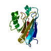
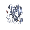
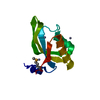
- PDB-3hgb: Crystal structure of glycine cleavage system protein H from Mycob... -
+ Open data

- Basic information
| Entry | Database: PDB / ID: 3hgb | ||||||
|---|---|---|---|---|---|---|---|
| Title | Crystal structure of glycine cleavage system protein H from Mycobacterium tuberculosis | ||||||
 Components Components | Glycine cleavage system H protein | ||||||
 Keywords Keywords | OXIDOREDUCTASE / SSGCID / NIAID / deCODE / UW / SBRI / Lipoyl / Structural Genomics / Seattle Structural Genomics Center for Infectious Disease / UNKNOWN FUNCTION | ||||||
| Function / homology |  Function and homology information Function and homology informationglycine cleavage complex / glycine decarboxylation via glycine cleavage system / cytoplasm / cytosol Similarity search - Function | ||||||
| Biological species |   Mycobacterium tuberculosis (bacteria) Mycobacterium tuberculosis (bacteria) | ||||||
| Method |  X-RAY DIFFRACTION / MOLECULAR REPLACEMENT / molecular replacement / Resolution: 1.75 Å X-RAY DIFFRACTION / MOLECULAR REPLACEMENT / molecular replacement / Resolution: 1.75 Å | ||||||
 Authors Authors | Seattle Structural Genomics Center for Infectious Disease (SSGCID) | ||||||
 Citation Citation | Journal: J.Struct.Funct.Genom. / Year: 2010 Title: X-ray structure determination of the glycine cleavage system protein H of Mycobacterium tuberculosis using an inverse Compton synchrotron X-ray source. Authors: Abendroth, J. / McCormick, M.S. / Edwards, T.E. / Staker, B. / Loewen, R. / Gifford, M. / Rifkin, J. / Mayer, C. / Guo, W. / Zhang, Y. / Myler, P. / Kelley, A. / Analau, E. / Hewitt, S.N. / ...Authors: Abendroth, J. / McCormick, M.S. / Edwards, T.E. / Staker, B. / Loewen, R. / Gifford, M. / Rifkin, J. / Mayer, C. / Guo, W. / Zhang, Y. / Myler, P. / Kelley, A. / Analau, E. / Hewitt, S.N. / Napuli, A.J. / Kuhn, P. / Ruth, R.D. / Stewart, L.J. #1: Journal: Tuberculosis (Edinb) / Year: 2015Title: Increasing the structural coverage of tuberculosis drug targets. Authors: Baugh, L. / Phan, I. / Begley, D.W. / Clifton, M.C. / Armour, B. / Dranow, D.M. / Taylor, B.M. / Muruthi, M.M. / Abendroth, J. / Fairman, J.W. / Fox, D. / Dieterich, S.H. / Staker, B.L. / ...Authors: Baugh, L. / Phan, I. / Begley, D.W. / Clifton, M.C. / Armour, B. / Dranow, D.M. / Taylor, B.M. / Muruthi, M.M. / Abendroth, J. / Fairman, J.W. / Fox, D. / Dieterich, S.H. / Staker, B.L. / Gardberg, A.S. / Choi, R. / Hewitt, S.N. / Napuli, A.J. / Myers, J. / Barrett, L.K. / Zhang, Y. / Ferrell, M. / Mundt, E. / Thompkins, K. / Tran, N. / Lyons-Abbott, S. / Abramov, A. / Sekar, A. / Serbzhinskiy, D. / Lorimer, D. / Buchko, G.W. / Stacy, R. / Stewart, L.J. / Edwards, T.E. / Van Voorhis, W.C. / Myler, P.J. | ||||||
| History |
|
- Structure visualization
| Structure viewer | Molecule: MolmilJmol/JSmol |
|---|
- Downloads & links
-Download
| PDBx/mmCIF format | 3hgb.cif.gz | 44.4 KB | Display | PDBx/mmCIF format |
|---|---|---|---|---|
| PDB format | pdb3hgb.ent.gz | 29.5 KB | Display | PDB format |
| PDBx/mmJSON format | 3hgb.json.gz | Tree view | PDBx/mmJSON format | |
| Others |  Other downloads Other downloads |
-Validation report
| Summary document | 3hgb_validation.pdf.gz | 422.2 KB | Display | wwPDB validaton report |
|---|---|---|---|---|
| Full document | 3hgb_full_validation.pdf.gz | 423.9 KB | Display | |
| Data in XML | 3hgb_validation.xml.gz | 9.2 KB | Display | |
| Data in CIF | 3hgb_validation.cif.gz | 13 KB | Display | |
| Arichive directory | https://data.pdbj.org/pub/pdb/validation_reports/hg/3hgbftp://data.pdbj.org/pub/pdb/validation_reports/hg/3hgb | HTTPS FTP |
-Related structure data
| Related structure data |  3iftC  1onlS S: Starting model for refinement C: citing same article ( |
|---|---|
| Similar structure data | |
| Other databases |


-Links
 PDBj
PDBj

- Assembly
| Deposited unit | 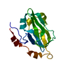
| ||||||||
|---|---|---|---|---|---|---|---|---|---|
| 1 |
| ||||||||
| Unit cell |
|
-Components
| #1: Protein | Mass: 16535.014 Da / Num. of mol.: 1 Source method: isolated from a genetically manipulated source Details: Expressed with an N-terminal hexahis tag and 3C cleavage site, but not cleaved prior to crystallization Source: (gene. exp.) Mycobacterium tuberculosis (bacteria) / Strain: H37Rv / Gene: gcvH, Rv1826, MT1874, MTCY1A11.17c / Plasmid: AVA0421 / Production host: |
|---|---|
| #2: Water | ChemComp-HOH /  Mass: 18.015 Da / Num. of mol.: 190 / Source method: isolated from a natural source / Formula: H2O Mass: 18.015 Da / Num. of mol.: 190 / Source method: isolated from a natural source / Formula: H2O |
-Experimental details
-Experiment
| Experiment | Method: X-RAY DIFFRACTION / Number of used crystals: 1 |
|---|
- Sample preparation
| Crystal | Density Matthews: 2.19 Å3/Da / Density % sol: 43.88 % |
|---|---|
| Crystal grow | Temperature: 289 K / Method: vapor diffusion, sitting drop / pH: 7.5 Details: JCSG+ screen condition B11, 1.6M Na citrate, 27.2 mg/mL protein, tracking ID 202071b11, pH 7.5, VAPOR DIFFUSION, SITTING DROP, temperature 289K |
-Data collection
| Diffraction | Mean temperature: 100 K |
|---|---|
| Diffraction source | Source: ROTATING ANODE / Type: RIGAKU FR-E+ SUPERBRIGHT / Wavelength: 1.5418 Å |
| Detector | Type: RIGAKU SATURN 944+ / Detector: CCD / Date: May 7, 2009 |
| Radiation | Protocol: SINGLE WAVELENGTH / Monochromatic (M) / Laue (L): M / Scattering type: x-ray |
| Radiation wavelength | Wavelength: 1.5418 Å / Relative weight: 1 |
| Reflection | Resolution: 1.75→19.24 Å / Num. obs: 14091 / % possible obs: 96.9 % / Observed criterion σ(I): -3 / Biso Wilson estimate: 17.299 Å2 / Rmerge(I) obs: 0.027 / Net I/σ(I): 23.75 |
| Reflection shell | Resolution: 1.75→1.8 Å / Rmerge(I) obs: 0.088 / Mean I/σ(I) obs: 6.2 / Num. measured obs: 1516 / Num. unique all: 1067 / Num. unique obs: 1067 / % possible all: 98.1 |
-Phasing
| Phasing | Method: molecular replacement | |||||||||
|---|---|---|---|---|---|---|---|---|---|---|
| Phasing MR |
|
- Processing
| Software |
| |||||||||||||||||||||||||||||||||||||||||||||||||||||||||||||||||
|---|---|---|---|---|---|---|---|---|---|---|---|---|---|---|---|---|---|---|---|---|---|---|---|---|---|---|---|---|---|---|---|---|---|---|---|---|---|---|---|---|---|---|---|---|---|---|---|---|---|---|---|---|---|---|---|---|---|---|---|---|---|---|---|---|---|---|
| Refinement | Method to determine structure: MOLECULAR REPLACEMENT Starting model: 1onl Resolution: 1.75→19.24 Å / Cor.coef. Fo:Fc: 0.943 / Cor.coef. Fo:Fc free: 0.919 / WRfactor Rfree: 0.206 / WRfactor Rwork: 0.169 / Occupancy max: 1 / Occupancy min: 0.3 / FOM work R set: 0.856 / SU B: 2.361 / SU ML: 0.076 / SU R Cruickshank DPI: 0.122 / SU Rfree: 0.119 / Cross valid method: THROUGHOUT / σ(F): 0 / ESU R: 0.122 / ESU R Free: 0.119 / Stereochemistry target values: MAXIMUM LIKELIHOOD Details: HYDROGENS HAVE BEEN ADDED IN THE RIDING POSITIONS; U VALUES: REFINED INDIVIDUALLY
| |||||||||||||||||||||||||||||||||||||||||||||||||||||||||||||||||
| Solvent computation | Ion probe radii: 0.8 Å / Shrinkage radii: 0.8 Å / VDW probe radii: 1.4 Å / Solvent model: MASK | |||||||||||||||||||||||||||||||||||||||||||||||||||||||||||||||||
| Displacement parameters | Biso max: 45.26 Å2 / Biso mean: 12.298 Å2 / Biso min: 3.01 Å2
| |||||||||||||||||||||||||||||||||||||||||||||||||||||||||||||||||
| Refinement step | Cycle: LAST / Resolution: 1.75→19.24 Å
| |||||||||||||||||||||||||||||||||||||||||||||||||||||||||||||||||
| Refine LS restraints |
| |||||||||||||||||||||||||||||||||||||||||||||||||||||||||||||||||
| LS refinement shell | Resolution: 1.75→1.795 Å / Total num. of bins used: 20
|
