 Movie
Movie Controller


+ Open data

- Basic information
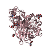
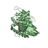
| Entry | Database: PDB / ID: 5wde | |||||||||
|---|---|---|---|---|---|---|---|---|---|---|
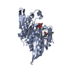
| Title | Crystal structure of the KIFC3 motor domain in complex with ADP | |||||||||
 Components Components | Kinesin-like protein KIFC3 | |||||||||
 Keywords Keywords |  MOTOR PROTEIN / kinesin / motor domain / adp / STRUCTURAL GENOMICS / STRUCTUR AL GENOMICS CONSORTIUM / SGC / Structural Genomics Consortium MOTOR PROTEIN / kinesin / motor domain / adp / STRUCTURAL GENOMICS / STRUCTUR AL GENOMICS CONSORTIUM / SGC / Structural Genomics Consortium | |||||||||
| Function / homology |  Function and homology informationzonula adherens maintenance / epithelial cell-cell adhesion / zonula adherens / minus-end-directed microtubule motor activity / kinesin complex / microtubule motor activity / microtubule-based movement / Golgi organization / Association of TriC/CCT with target proteins during biosynthesis / visual perception ...zonula adherens maintenance / epithelial cell-cell adhesion / zonula adherens / minus-end-directed microtubule motor activity / kinesin complex / microtubule motor activity / microtubule-based movement / Golgi organization / Association of TriC/CCT with target proteins during biosynthesis / visual perception / cytoplasmic vesicle membrane / microtubule binding / microtubule / centrosome / Golgi apparatus / extracellular exosome / ATP binding / identical protein binding Function and homology informationzonula adherens maintenance / epithelial cell-cell adhesion / zonula adherens / minus-end-directed microtubule motor activity / kinesin complex / microtubule motor activity / microtubule-based movement / Golgi organization / Association of TriC/CCT with target proteins during biosynthesis / visual perception ...zonula adherens maintenance / epithelial cell-cell adhesion / zonula adherens / minus-end-directed microtubule motor activity / kinesin complex / microtubule motor activity / microtubule-based movement / Golgi organization / Association of TriC/CCT with target proteins during biosynthesis / visual perception / cytoplasmic vesicle membrane / microtubule binding / microtubule / centrosome / Golgi apparatus / extracellular exosome / ATP binding / identical protein bindingSimilarity search - Function | |||||||||
| Biological species |  Homo sapiens (human) Homo sapiens (human) | |||||||||
| Method | X-RAY DIFFRACTION / SYNCHROTRON / MOLECULAR REPLACEMENT / Resolution: 1.85 Å | |||||||||
 Authors Authors | Shen, Y. / Tempel, W. / Landry, R. / Arrowsmith, C.H. / Edwards, A.M. / Park, H. / Structural Genomics Consortium (SGC) | |||||||||
 Citation Citation | Journal: Sci Rep / Year: 2017 Title: Structural basis of small molecule ATPase inhibition of a human mitotic kinesin motor protein. Authors: Park, H.W. / Ma, Z. / Zhu, H. / Jiang, S. / Robinson, R.C. / Endow, S.A. | |||||||||
| History |
|
- Structure visualization
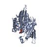
| Structure viewer | Molecule: MolmilJmol/JSmol |
|---|
- Downloads & links
-Download
| PDBx/mmCIF format | 5wde.cif.gz | 147 KB | Display | PDBx/mmCIF format |
|---|---|---|---|---|
| PDB format | pdb5wde.ent.gz | 112.3 KB | Display | PDB format |
| PDBx/mmJSON format | 5wde.json.gz | Tree view | PDBx/mmJSON format | |
| Others |  Other downloads Other downloads |
-Validation report
| Arichive directory | https://data.pdbj.org/pub/pdb/validation_reports/wd/5wdeftp://data.pdbj.org/pub/pdb/validation_reports/wd/5wde | HTTPS FTP |
|---|
-Related structure data
| Related structure data | 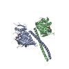 5w3dC 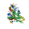 5wdhC 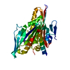 1f9tS S: Starting model for refinement C: citing same article ( |
|---|---|
| Similar structure data |


-Links
 PDBj
PDBj


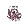
- Assembly
| Deposited unit | 
| ||||||||
|---|---|---|---|---|---|---|---|---|---|
| 1 |
| ||||||||
| Unit cell |
| ||||||||
| Details | Author states that there is no direct experimental evidence for biological assembly since this is a fragment of full length protein. |
-Components
| #1: Protein | Mass: 36221.527 Da / Num. of mol.: 1 / Fragment: motor domain (UNP residues 443-770) Source method: isolated from a genetically manipulated source Source: (gene. exp.) Homo sapiens (human) / Gene: KIFC3 / Plasmid: p28a-LIC / Production host:  Escherichia coli (E. coli) / Strain (production host): BL21-CodonPlus (DE-3)-RIL / References: UniProt: Q9BVG8 Escherichia coli (E. coli) / Strain (production host): BL21-CodonPlus (DE-3)-RIL / References: UniProt: Q9BVG8 | ||
|---|---|---|---|
| #2: Chemical | ChemComp-ADP / Adenosine diphosphate  Mass: 427.201 Da / Num. of mol.: 1 / Source method: obtained synthetically / Formula: C10H15N5O10P2 / Comment: ADP, energy-carrying molecule*YM Mass: 427.201 Da / Num. of mol.: 1 / Source method: obtained synthetically / Formula: C10H15N5O10P2 / Comment: ADP, energy-carrying molecule*YM | ||
| #3: Chemical | ChemComp-MG /   Mass: 24.305 Da / Num. of mol.: 1 / Source method: obtained synthetically / Formula: Mg Mass: 24.305 Da / Num. of mol.: 1 / Source method: obtained synthetically / Formula: Mg | ||
| #4: Chemical | ChemComp-UNX /   Num. of mol.: 46 / Source method: obtained synthetically Num. of mol.: 46 / Source method: obtained synthetically#5: Water | ChemComp-HOH / | Water Mass: 18.015 Da / Num. of mol.: 126 / Source method: isolated from a natural source / Formula: H2O Mass: 18.015 Da / Num. of mol.: 126 / Source method: isolated from a natural source / Formula: H2O |
-Experimental details
-Experiment
| Experiment | Method: X-RAY DIFFRACTION / Number of used crystals: 1 |
|---|
- Sample preparation
| Crystal | Density Matthews: 7.3 Å3/Da / Density % sol: 83.1 % |
|---|---|
| Crystal grow | Temperature: 291 K / Method: vapor diffusion, sitting drop / pH: 7.5 / Details: 1.2M sodium citrate, 0.1M HEPES |
-Data collection
| Diffraction | Mean temperature: 100 K | ||||||||||||||||||||||||||||||
|---|---|---|---|---|---|---|---|---|---|---|---|---|---|---|---|---|---|---|---|---|---|---|---|---|---|---|---|---|---|---|---|
| Diffraction source | Source: SYNCHROTRON / Site: CHESS  / Beamline: F1 / Wavelength: 0.9176 Å / Beamline: F1 / Wavelength: 0.9176 Å | ||||||||||||||||||||||||||||||
| Detector | Type: ADSC QUANTUM 4 / Detector: CCD / Date: May 7, 2006 | ||||||||||||||||||||||||||||||
| Radiation | Protocol: SINGLE WAVELENGTH / Monochromatic (M) / Laue (L): M / Scattering type: x-ray | ||||||||||||||||||||||||||||||
| Radiation wavelength | Wavelength: 0.9176 Å / Relative weight: 1 | ||||||||||||||||||||||||||||||
| Reflection | Resolution: 1.85→47.69 Å / Num. obs: 86307 / % possible obs: 99.7 % / Redundancy: 6.4 % / Biso Wilson estimate: 29.73 Å2 / CC1/2: 1 / Rmerge(I) obs: 0.048 / Rpim(I) all: 0.02 / Rrim(I) all: 0.052 / Net I/σ(I): 22.2 / Num. measured all: 549983 / Scaling rejects: 0 | ||||||||||||||||||||||||||||||
| Reflection shell | Diffraction-ID: 1
|
- Processing
| Software |
| |||||||||||||||||||||||||||||||||||||||||||||||||||||||||||||||||||||||||||||||||||||||||||||||||||||||||||||||||||||||||||||||||||||||||||||||||||||||||||||||||||||||||||||||||||||||||||||||||||||||||||||||||||||||||||||||||
|---|---|---|---|---|---|---|---|---|---|---|---|---|---|---|---|---|---|---|---|---|---|---|---|---|---|---|---|---|---|---|---|---|---|---|---|---|---|---|---|---|---|---|---|---|---|---|---|---|---|---|---|---|---|---|---|---|---|---|---|---|---|---|---|---|---|---|---|---|---|---|---|---|---|---|---|---|---|---|---|---|---|---|---|---|---|---|---|---|---|---|---|---|---|---|---|---|---|---|---|---|---|---|---|---|---|---|---|---|---|---|---|---|---|---|---|---|---|---|---|---|---|---|---|---|---|---|---|---|---|---|---|---|---|---|---|---|---|---|---|---|---|---|---|---|---|---|---|---|---|---|---|---|---|---|---|---|---|---|---|---|---|---|---|---|---|---|---|---|---|---|---|---|---|---|---|---|---|---|---|---|---|---|---|---|---|---|---|---|---|---|---|---|---|---|---|---|---|---|---|---|---|---|---|---|---|---|---|---|---|---|---|---|---|---|---|---|---|---|---|---|---|---|---|---|---|---|
| Refinement | Method to determine structure: MOLECULAR REPLACEMENT Starting model: 1f9t Resolution: 1.85→27.079 Å / SU ML: 0.2 / Cross valid method: FREE R-VALUE / σ(F): 0.1 / Phase error: 24.68 Details: This REDO of the KIFC3 structure was in part motivated by the PDB-REDO model of PDB entry 2H58. Diffraction images that formed the basis for PDB entry 2H58 were reprocessed. Merging ...Details: This REDO of the KIFC3 structure was in part motivated by the PDB-REDO model of PDB entry 2H58. Diffraction images that formed the basis for PDB entry 2H58 were reprocessed. Merging statistics indicate significant anisotropy of diffraction. For this round of model refinement, new crossvalidation flags were assigned to the reflections using the CCP4 FREERFLAG command. This reassignment was followed by torsion angle simulated annealing. We note the high solvent content of this crystal form and the presence of many uninterpreted features in the difference maps when contoured at 1.5*rmsd (2FOFCWT) or +/-3*rmsd (FOFCWT). We thank CCP4BB participants who responded to questions related to this structure on the board and off-list.
| |||||||||||||||||||||||||||||||||||||||||||||||||||||||||||||||||||||||||||||||||||||||||||||||||||||||||||||||||||||||||||||||||||||||||||||||||||||||||||||||||||||||||||||||||||||||||||||||||||||||||||||||||||||||||||||||||
| Solvent computation | Shrinkage radii: 0.9 Å / VDW probe radii: 1.11 Å | |||||||||||||||||||||||||||||||||||||||||||||||||||||||||||||||||||||||||||||||||||||||||||||||||||||||||||||||||||||||||||||||||||||||||||||||||||||||||||||||||||||||||||||||||||||||||||||||||||||||||||||||||||||||||||||||||
| Displacement parameters | Biso max: 95.04 Å2 / Biso mean: 37.5357 Å2 / Biso min: 20.74 Å2 | |||||||||||||||||||||||||||||||||||||||||||||||||||||||||||||||||||||||||||||||||||||||||||||||||||||||||||||||||||||||||||||||||||||||||||||||||||||||||||||||||||||||||||||||||||||||||||||||||||||||||||||||||||||||||||||||||
| Refinement step | Cycle: final / Resolution: 1.85→27.079 Å
| |||||||||||||||||||||||||||||||||||||||||||||||||||||||||||||||||||||||||||||||||||||||||||||||||||||||||||||||||||||||||||||||||||||||||||||||||||||||||||||||||||||||||||||||||||||||||||||||||||||||||||||||||||||||||||||||||
| Refine LS restraints |
| |||||||||||||||||||||||||||||||||||||||||||||||||||||||||||||||||||||||||||||||||||||||||||||||||||||||||||||||||||||||||||||||||||||||||||||||||||||||||||||||||||||||||||||||||||||||||||||||||||||||||||||||||||||||||||||||||
| LS refinement shell | Refine-ID: X-RAY DIFFRACTION / Total num. of bins used: 30
| |||||||||||||||||||||||||||||||||||||||||||||||||||||||||||||||||||||||||||||||||||||||||||||||||||||||||||||||||||||||||||||||||||||||||||||||||||||||||||||||||||||||||||||||||||||||||||||||||||||||||||||||||||||||||||||||||
| Refinement TLS params. | Method: refined / Refine-ID: X-RAY DIFFRACTION
| |||||||||||||||||||||||||||||||||||||||||||||||||||||||||||||||||||||||||||||||||||||||||||||||||||||||||||||||||||||||||||||||||||||||||||||||||||||||||||||||||||||||||||||||||||||||||||||||||||||||||||||||||||||||||||||||||
| Refinement TLS group |
|
