 Movie
Movie Controller

[English] 日本語
 Yorodumi
- PDB-5wcj: Crystal Structure of Human Methyltransferase-like protein 13 in c... -
+ Open data

- Basic information
| Entry | Database: PDB / ID: 5wcj | ||||||
|---|---|---|---|---|---|---|---|
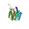
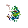
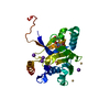
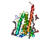
| Title | Crystal Structure of Human Methyltransferase-like protein 13 in complex with SAH | ||||||
 Components Components | Methyltransferase-like protein 13 | ||||||
 Keywords Keywords |  TRANSFERASE / methyltransferase / Rossman fold / SAH-binding / SGC / Structural Genomics / Structural Genomics Consortium TRANSFERASE / methyltransferase / Rossman fold / SAH-binding / SGC / Structural Genomics / Structural Genomics Consortium | ||||||
| Function / homology |  Function and homology information Function and homology informationnegative regulation of cell cycle G1/S phase transition / Transferases; Transferring one-carbon groups; Methyltransferases / methyltransferase activity / methylation / negative regulation of transcription by RNA polymerase II / mitochondrion / nucleus / cytoplasmSimilarity search - Function | ||||||
| Biological species |  Homo sapiens (human) Homo sapiens (human) | ||||||
| Method | X-RAY DIFFRACTION / SYNCHROTRON / MOLECULAR REPLACEMENT / Resolution: 1.7 Å | ||||||
 Authors Authors | Halabelian, L. / Loppnau, P. / Seitova, A. / Hutchinson, A. / Hunt, B. / Dong, A. / Bountra, C. / Edwards, A.M. / Arrowsmith, C.H. / Structural Genomics Consortium (SGC) | ||||||
 Citation Citation | Journal: Nat Commun / Year: 2018 Title: The dual methyltransferase METTL13 targets N terminus and Lys55 of eEF1A and modulates codon-specific translation rates. Authors: Jakobsson, M.E. / Malecki, J.M. / Halabelian, L. / Nilges, B.S. / Pinto, R. / Kudithipudi, S. / Munk, S. / Davydova, E. / Zuhairi, F.R. / Arrowsmith, C.H. / Jeltsch, A. / Leidel, S.A. / ...Authors: Jakobsson, M.E. / Malecki, J.M. / Halabelian, L. / Nilges, B.S. / Pinto, R. / Kudithipudi, S. / Munk, S. / Davydova, E. / Zuhairi, F.R. / Arrowsmith, C.H. / Jeltsch, A. / Leidel, S.A. / Olsen, J.V. / Falnes, P.O. | ||||||
| History |
|
- Structure visualization
| Structure viewer | Molecule: MolmilJmol/JSmol |
|---|
- Downloads & links
-Download
| PDBx/mmCIF format | 5wcj.cif.gz | 61.3 KB | Display | PDBx/mmCIF format |
|---|---|---|---|---|
| PDB format | pdb5wcj.ent.gz | 41.3 KB | Display | PDB format |
| PDBx/mmJSON format | 5wcj.json.gz | Tree view | PDBx/mmJSON format | |
| Others |  Other downloads Other downloads |
-Validation report
| Arichive directory | https://data.pdbj.org/pub/pdb/validation_reports/wc/5wcjftp://data.pdbj.org/pub/pdb/validation_reports/wc/5wcj | HTTPS FTP |
|---|
-Related structure data
| Related structure data |  3gjyS S: Starting model for refinement |
|---|---|
| Similar structure data |


-Links
 PDBj
PDBj- Assembly
| Deposited unit | 
| ||||||||
|---|---|---|---|---|---|---|---|---|---|
| 1 |
| ||||||||
| Unit cell |
|
-Components
| #1: Protein | Mass: 27539.996 Da / Num. of mol.: 1 Source method: isolated from a genetically manipulated source Source: (gene. exp.) Homo sapiens (human) / Gene: METTL13, KIAA0859, CGI-01 / Plasmid: pFBOH-MHL / Production host:   Spodoptera frugiperda (fall armyworm) / Strain (production host): SF9 cells Spodoptera frugiperda (fall armyworm) / Strain (production host): SF9 cellsReferences: UniProt: Q8N6R0, Transferases; Transferring one-carbon groups; Methyltransferases | ||||
|---|---|---|---|---|---|
| #2: Chemical | ChemComp-UNX /   Num. of mol.: 4 / Source method: obtained synthetically Num. of mol.: 4 / Source method: obtained synthetically#3: Chemical | ChemComp-SAH / | S-Adenosyl-L-homocysteine  Type: L-peptide linking / Mass: 384.411 Da / Num. of mol.: 1 / Source method: obtained synthetically / Formula: C14H20N6O5S Type: L-peptide linking / Mass: 384.411 Da / Num. of mol.: 1 / Source method: obtained synthetically / Formula: C14H20N6O5S#4: Water | ChemComp-HOH / | Water Mass: 18.015 Da / Num. of mol.: 62 / Source method: isolated from a natural source / Formula: H2O Mass: 18.015 Da / Num. of mol.: 62 / Source method: isolated from a natural source / Formula: H2O |
-Experimental details
-Experiment
| Experiment | Method: X-RAY DIFFRACTION / Number of used crystals: 1 |
|---|
- Sample preparation
| Crystal | Density Matthews: 2.19 Å3/Da / Density % sol: 43.74 % / Mosaicity: 0.21 ° |
|---|---|
| Crystal grow | Temperature: 291 K / Method: vapor diffusion, sitting drop / pH: 7.4 / Details: 20% PEG3350, 200mM Ammonium Chloride |
-Data collection
| Diffraction | Mean temperature: 100 K | ||||||||||||||||||||||||||||||
|---|---|---|---|---|---|---|---|---|---|---|---|---|---|---|---|---|---|---|---|---|---|---|---|---|---|---|---|---|---|---|---|
| Diffraction source | Source: SYNCHROTRON / Site: APS  / Beamline: 19-ID / Wavelength: 0.97863 Å / Beamline: 19-ID / Wavelength: 0.97863 Å | ||||||||||||||||||||||||||||||
| Detector | Type: DECTRIS PILATUS3 6M / Detector: PIXEL / Date: Jun 23, 2017 | ||||||||||||||||||||||||||||||
| Radiation | Protocol: SINGLE WAVELENGTH / Monochromatic (M) / Laue (L): M / Scattering type: x-ray | ||||||||||||||||||||||||||||||
| Radiation wavelength | Wavelength: 0.97863 Å / Relative weight: 1 | ||||||||||||||||||||||||||||||
| Reflection | Resolution: 1.7→35 Å / Num. obs: 26945 / % possible obs: 99 % / Redundancy: 6.4 % / CC1/2: 0.999 / Rmerge(I) obs: 0.053 / Rpim(I) all: 0.023 / Rrim(I) all: 0.058 / Net I/σ(I): 17.5 / Num. measured all: 172955 / Scaling rejects: 0 | ||||||||||||||||||||||||||||||
| Reflection shell | Diffraction-ID: 1
|
- Processing
| Software |
| ||||||||||||||||||||||||||||||||||||||||||||||||||||||||||||
|---|---|---|---|---|---|---|---|---|---|---|---|---|---|---|---|---|---|---|---|---|---|---|---|---|---|---|---|---|---|---|---|---|---|---|---|---|---|---|---|---|---|---|---|---|---|---|---|---|---|---|---|---|---|---|---|---|---|---|---|---|---|
| Refinement | Method to determine structure: MOLECULAR REPLACEMENT Starting model: 3GJY Resolution: 1.7→35 Å / Cor.coef. Fo:Fc: 0.968 / Cor.coef. Fo:Fc free: 0.958 / SU B: 2.61 / SU ML: 0.08 / Cross valid method: THROUGHOUT / σ(F): 0 / ESU R: 0.1 / ESU R Free: 0.097 Details: HYDROGENS HAVE BEEN ADDED IN THE RIDING POSITIONS U VALUES : REFINED INDIVIDUALLY
| ||||||||||||||||||||||||||||||||||||||||||||||||||||||||||||
| Solvent computation | Ion probe radii: 0.8 Å / Shrinkage radii: 0.8 Å / VDW probe radii: 1.2 Å | ||||||||||||||||||||||||||||||||||||||||||||||||||||||||||||
| Displacement parameters | Biso max: 93.48 Å2 / Biso mean: 35.246 Å2 / Biso min: 20.57 Å2
| ||||||||||||||||||||||||||||||||||||||||||||||||||||||||||||
| Refinement step | Cycle: final / Resolution: 1.7→35 Å
| ||||||||||||||||||||||||||||||||||||||||||||||||||||||||||||
| Refine LS restraints |
| ||||||||||||||||||||||||||||||||||||||||||||||||||||||||||||
| LS refinement shell | Resolution: 1.7→1.744 Å / Rfactor Rfree error: 0 / Total num. of bins used: 20
|
