 Movie
Movie Controller


+ Open data

- Basic information
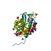
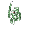
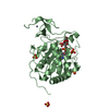
| Entry | Database: PDB / ID: 4mat | ||||||
|---|---|---|---|---|---|---|---|
| Title | E.COLI METHIONINE AMINOPEPTIDASE HIS79ALA MUTANT | ||||||
 Components Components | PROTEIN (METHIONINE AMINOPEPTIDASE) | ||||||
 Keywords Keywords |  HYDROLASE / HYDROLASE(ALPHA-AMINOACYLPEPTIDE) / SITE-DIRECTED MUTANT HYDROLASE / HYDROLASE(ALPHA-AMINOACYLPEPTIDE) / SITE-DIRECTED MUTANT | ||||||
| Function / homology |  Function and homology information Function and homology information: / initiator methionyl aminopeptidase activity / methionyl aminopeptidase / metalloaminopeptidase activity / ferrous iron binding / proteolysis / metal ion binding / cytosolSimilarity search - Function | ||||||
| Biological species |  Escherichia coli (E. coli) Escherichia coli (E. coli) | ||||||
| Method | X-RAY DIFFRACTION / MOLECULAR REPLACEMENT / Resolution: 2 Å | ||||||
 Authors Authors | Lowther, W.T. / Orville, A.M. / Madden, D.T. / Lim, S. / Rich, D.H. / Matthews, B.W. | ||||||
 Citation Citation | Journal: Biochemistry / Year: 1999 Title: Escherichia coli methionine aminopeptidase: implications of crystallographic analyses of the native, mutant, and inhibited enzymes for the mechanism of catalysis. Authors: Lowther, W.T. / Orville, A.M. / Madden, D.T. / Lim, S. / Rich, D.H. / Matthews, B.W. #1: Journal: Proc.Natl.Acad.Sci.USA / Year: 1998 Title: The anti-angiogenic agent fumagillin covalently modifies a conserved active-site histidine in the Escherichia coli methionine aminopeptidase. Authors: Lowther, W.T. / McMillen, D.A. / Orville, A.M. / Matthews, B.W. #2: Journal: Biochemistry / Year: 1993Title: Structure of the cobalt-dependent methionine aminopeptidase from Escherichia coli: a new type of proteolytic enzyme. Authors: Roderick, S.L. / Matthews, B.W. | ||||||
| History |
|
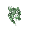
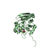
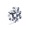
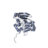
- Structure visualization
| Structure viewer | Molecule: MolmilJmol/JSmol |
|---|
- Downloads & links
-Download
| PDBx/mmCIF format | 4mat.cif.gz | 69.7 KB | Display | PDBx/mmCIF format |
|---|---|---|---|---|
| PDB format | pdb4mat.ent.gz | 49.7 KB | Display | PDB format |
| PDBx/mmJSON format | 4mat.json.gz | Tree view | PDBx/mmJSON format | |
| Others |  Other downloads Other downloads |
-Validation report
| Arichive directory | https://data.pdbj.org/pub/pdb/validation_reports/ma/4matftp://data.pdbj.org/pub/pdb/validation_reports/ma/4mat | HTTPS FTP |
|---|
-Related structure data
| Related structure data | 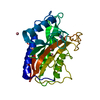 2matSC 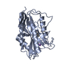 3matC S: Starting model for refinement C: citing same article ( |
|---|---|
| Similar structure data |


-Links
 PDBj
PDBj- Assembly
| Deposited unit | 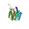
| ||||||||
|---|---|---|---|---|---|---|---|---|---|
| 1 |
| ||||||||
| Unit cell |
|
-Components
| #1: Protein | Mass: 31023.641 Da / Num. of mol.: 1 / Mutation: R175Q, H79A Source method: isolated from a genetically manipulated source Details: SITE-DIRECTED MUTANT / Source: (gene. exp.) Escherichia coli (E. coli) / Plasmid: PET28B / Cell line (production host): BL21(DE3) / Production host: Escherichia coli (E. coli)References: UniProt: P07906, UniProt: P0AE18*PLUS, methionyl aminopeptidase |
|---|---|
| #2: Chemical | ChemComp-NA /   Mass: 22.990 Da / Num. of mol.: 1 / Source method: obtained synthetically / Formula: Na Mass: 22.990 Da / Num. of mol.: 1 / Source method: obtained synthetically / Formula: Na |
| #3: Water | ChemComp-HOH / Water Mass: 18.015 Da / Num. of mol.: 155 / Source method: isolated from a natural source / Formula: H2O Mass: 18.015 Da / Num. of mol.: 155 / Source method: isolated from a natural source / Formula: H2O |
| Sequence details | POLY-HISTIDINE TAGGED VARIANT THIS PROTEIN WAS NOT TREATED WITH THROMBIN AND THEREFORE CONTAINED ...POLY-HISTIDINE TAGGED VARIANT THIS PROTEIN WAS NOT TREATED WITH THROMBIN AND THEREFORE CONTAINED ADDITIONAL |
-Experimental details
-Experiment
| Experiment | Method: X-RAY DIFFRACTION / Number of used crystals: 1 |
|---|
- Sample preparation
| Crystal | Density Matthews: 2.38 Å3/Da / Density % sol: 47.9 % | ||||||||||||||||||||||||||||||||||||
|---|---|---|---|---|---|---|---|---|---|---|---|---|---|---|---|---|---|---|---|---|---|---|---|---|---|---|---|---|---|---|---|---|---|---|---|---|---|
| Crystal grow | pH: 7 Details: Crystals of the HIS79ALA mutant were obtained by mixing the apoenzyme retaining the C-terminal HISs-tag (6.7 mg/ml, 20 mM DTT, 25 mM HEPES pH 6.8, 25 mM K2SO4, 100 mM NaCl) with an equal ...Details: Crystals of the HIS79ALA mutant were obtained by mixing the apoenzyme retaining the C-terminal HISs-tag (6.7 mg/ml, 20 mM DTT, 25 mM HEPES pH 6.8, 25 mM K2SO4, 100 mM NaCl) with an equal volume of well solution (22-27 % PEG 3400, 0.1 M HEPES pH 7.0, 200 mM NaCl). Diffraction quality crystals were obtained after macroseeding into 20 uL hanging drops. | ||||||||||||||||||||||||||||||||||||
| Crystal grow | *PLUS Method: vapor diffusion, hanging drop / Details: macroseeding | ||||||||||||||||||||||||||||||||||||
| Components of the solutions | *PLUS
|
-Data collection
| Diffraction | Mean temperature: 103 K |
|---|---|
| Diffraction source | Source: ROTATING ANODE / Type: RIGAKU RU200 / Wavelength: 1.5418 |
| Detector | Type: RIGAKU RAXIS IV / Detector: IMAGE PLATE / Date: Oct 10, 1997 |
| Radiation | Monochromator: NI FILTER / Protocol: SINGLE WAVELENGTH / Monochromatic (M) / Laue (L): M / Scattering type: x-ray |
| Radiation wavelength | Wavelength: 1.5418 Å / Relative weight: 1 |
| Reflection | Resolution: 2→28.8 Å / Num. obs: 16062 / % possible obs: 85.2 % / Redundancy: 3 % / Biso Wilson estimate: 16.2 Å2 / Rsym value: 4.7 / Net I/σ(I): 22.4 |
| Reflection shell | Resolution: 2→2.07 Å / Mean I/σ(I) obs: 4.4 / Rsym value: 21 / % possible all: 70.8 |
| Reflection | *PLUS Num. measured all: 48052 / Rmerge(I) obs: 0.047 |
| Reflection shell | *PLUS % possible obs: 70.8 % / Rmerge(I) obs: 0.21 |
- Processing
| Software |
| ||||||||||||||||||||||||||||||||||||||||||||||||||
|---|---|---|---|---|---|---|---|---|---|---|---|---|---|---|---|---|---|---|---|---|---|---|---|---|---|---|---|---|---|---|---|---|---|---|---|---|---|---|---|---|---|---|---|---|---|---|---|---|---|---|---|
| Refinement | Method to determine structure: MOLECULAR REPLACEMENT Starting model: 2MAT Resolution: 2→28.8 Å / Isotropic thermal model: TNT BCORREL V1.0 / σ(F): 0 / Stereochemistry target values: TNT PROTGEO
| ||||||||||||||||||||||||||||||||||||||||||||||||||
| Solvent computation | Solvent model: TNT / Bsol: 207.2 Å2 / ksol: 0.915 e/Å3 | ||||||||||||||||||||||||||||||||||||||||||||||||||
| Refinement step | Cycle: LAST / Resolution: 2→28.8 Å
| ||||||||||||||||||||||||||||||||||||||||||||||||||
| Refine LS restraints |
| ||||||||||||||||||||||||||||||||||||||||||||||||||
| Software | *PLUS Name: TNT / Version: 5F / Classification: refinement | ||||||||||||||||||||||||||||||||||||||||||||||||||
| Refine LS restraints | *PLUS
|
