 Movie
Movie Controller

[English] 日本語
 Yorodumi
- PDB-5w59: Crystal structure of a monomeric human FGF9 in complex with the e... -
+ Open data

- Basic information
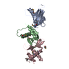
| Entry | Database: PDB / ID: 5w59 | ||||||
|---|---|---|---|---|---|---|---|
| Title | Crystal structure of a monomeric human FGF9 in complex with the ectodomain of human FGFR1c | ||||||
 Components Components |
| ||||||
 Keywords Keywords |  CYTOKINE / Growth factor / Growth Factor Receptor / ligand-receptor complex CYTOKINE / Growth factor / Growth Factor Receptor / ligand-receptor complex | ||||||
| Function / homology |  Function and homology information Function and homology informationregulation of timing of cell differentiation / positive regulation of activin receptor signaling pathway / negative regulation of vascular associated smooth muscle cell differentiation involved in phenotypic switching / positive regulation of reproductive process / Sertoli cell proliferation / Transcriptional regulation of testis differentiation / Signaling by FGFR1 amplification mutants / negative regulation of fibroblast growth factor production / positive regulation of mitotic cell cycle DNA replication / regulation of extrinsic apoptotic signaling pathway in absence of ligand ...regulation of timing of cell differentiation / positive regulation of activin receptor signaling pathway / negative regulation of vascular associated smooth muscle cell differentiation involved in phenotypic switching / positive regulation of reproductive process / Sertoli cell proliferation / Transcriptional regulation of testis differentiation / Signaling by FGFR1 amplification mutants / negative regulation of fibroblast growth factor production / positive regulation of mitotic cell cycle DNA replication / regulation of extrinsic apoptotic signaling pathway in absence of ligand / Signaling by plasma membrane FGFR1 fusions / diphosphate metabolic process / FGFR1c and Klotho ligand binding and activation / vitamin D3 metabolic process / regulation of phosphate transport / regulation of lateral mesodermal cell fate specification / positive regulation of MAPKKK cascade by fibroblast growth factor receptor signaling pathway / cementum mineralization / embryonic skeletal system development / response to sodium phosphate / FGFR3b ligand binding and activation / regulation of branching involved in salivary gland morphogenesis by mesenchymal-epithelial signaling / receptor-receptor interaction / fibroblast growth factor receptor signaling pathway involved in orbitofrontal cortex development / auditory receptor cell development / ventricular zone neuroblast division / Epithelial-Mesenchymal Transition (EMT) during gastrulation / eye development / positive regulation of parathyroid hormone secretion / chordate embryonic development / Signaling by activated point mutants of FGFR3 / FGFR3c ligand binding and activation / embryonic digestive tract development / Phospholipase C-mediated cascade; FGFR3 / mesenchymal cell proliferation / fibroblast growth factor receptor binding / paraxial mesoderm development / FGFR2c ligand binding and activation / Activated point mutants of FGFR2 / Phospholipase C-mediated cascade; FGFR2 / FGFR4 ligand binding and activation / FGFR1b ligand binding and activation / fibroblast growth factor receptor activity / branching involved in salivary gland morphogenesis / Phospholipase C-mediated cascade; FGFR4 / male sex determination / activin receptor signaling pathway / Signaling by activated point mutants of FGFR1 / FGFR1c ligand binding and activation / organ induction / Downstream signaling of activated FGFR1 / Phospholipase C-mediated cascade: FGFR1 / positive regulation of phospholipase activity / lung-associated mesenchyme development / cell projection assembly / positive regulation of smoothened signaling pathway / positive regulation of vascular associated smooth muscle cell migration / cellular response to fibroblast growth factor stimulus / positive regulation of vascular endothelial growth factor receptor signaling pathway / outer ear morphogenesis / middle ear morphogenesis / embryonic limb morphogenesis / skeletal system morphogenesis / positive regulation of endothelial cell chemotaxis / positive regulation of mesenchymal cell proliferation / cardiac muscle cell proliferation / positive regulation of vascular endothelial cell proliferation / ureteric bud development / smoothened signaling pathway / inner ear morphogenesis / midbrain development / PI-3K cascade:FGFR3 / negative regulation of Wnt signaling pathway / fibroblast growth factor binding / PI-3K cascade:FGFR2 / positive regulation of stem cell proliferation / PI-3K cascade:FGFR4 / Formation of paraxial mesoderm / PI-3K cascade:FGFR1 / regulation of cell differentiation / phosphatidylinositol-mediated signaling / positive regulation of cell division / PI3K Cascade / epithelial to mesenchymal transition / canonical Wnt signaling pathway / positive regulation of blood vessel endothelial cell migration / fibroblast growth factor receptor signaling pathway / calcium ion homeostasis / chondrocyte differentiation / SHC-mediated cascade:FGFR3 / vascular endothelial growth factor receptor signaling pathway / : / SHC-mediated cascade:FGFR2 / SHC-mediated cascade:FGFR4 / SHC-mediated cascade:FGFR1 / FRS-mediated FGFR3 signaling / cell maturation / positive regulation of cardiac muscle cell proliferation / FRS-mediated FGFR2 signaling / regulation of cell migrationSimilarity search - Function | ||||||
| Biological species |  Homo sapiens (human) Homo sapiens (human) | ||||||
| Method | X-RAY DIFFRACTION / SYNCHROTRON / MOLECULAR REPLACEMENT / Resolution: 2.498 Å | ||||||
 Authors Authors | Mohammadi, M. / Ma, J. / Liu, Y. | ||||||
| Funding support |  United States, 1items United States, 1items
| ||||||
 Citation Citation | Journal: Structure / Year: 2017 Title: Regulation of Receptor Binding Specificity of FGF9 by an Autoinhibitory Homodimerization. Authors: Liu, Y. / Ma, J. / Beenken, A. / Srinivasan, L. / Eliseenkova, A.V. / Mohammadi, M. | ||||||
| History |
|
- Structure visualization
| Structure viewer | Molecule: MolmilJmol/JSmol |
|---|
- Downloads & links
-Download
| PDBx/mmCIF format | 5w59.cif.gz | 160.6 KB | Display | PDBx/mmCIF format |
|---|---|---|---|---|
| PDB format | pdb5w59.ent.gz | 124.3 KB | Display | PDB format |
| PDBx/mmJSON format | 5w59.json.gz | Tree view | PDBx/mmJSON format | |
| Others |  Other downloads Other downloads |
-Validation report
| Arichive directory | https://data.pdbj.org/pub/pdb/validation_reports/w5/5w59ftp://data.pdbj.org/pub/pdb/validation_reports/w5/5w59 | HTTPS FTP |
|---|
-Related structure data
| Related structure data | 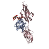 3ojvS S: Starting model for refinement |
|---|---|
| Similar structure data |


-Links
 PDBj
PDBj


- Assembly
| Deposited unit | 
| ||||||||
|---|---|---|---|---|---|---|---|---|---|
| 1 |
| ||||||||
| Unit cell |
|
-Components
| #1: Protein | / FGF-9 / Glia-activating factor / GAF / Heparin-binding growth factor 9 / HBGF-9 Mass: 19994.564 Da / Num. of mol.: 1 / Fragment: UNP residues 35-208 / Mutation: D195A/L204A/L205A Source method: isolated from a genetically manipulated source Details: Human FGF9 carrying D195A/L204A/L205A triple mutation in its C-terminal tail that renders the ligand monomeric Source: (gene. exp.) Homo sapiens (human) / Gene: FGF9 / Production host:  Escherichia coli (E. coli) / References: UniProt: P31371 Escherichia coli (E. coli) / References: UniProt: P31371 | ||
|---|---|---|---|
| #2: Protein | / FGFR-1 / Basic fibroblast growth factor receptor 1 / bFGF-R-1 / Fms-like tyrosine kinase 2 / FLT-2 ...FGFR-1 / Basic fibroblast growth factor receptor 1 / bFGF-R-1 / Fms-like tyrosine kinase 2 / FLT-2 / N-sam / Proto-oncogene c-Fgr Mass: 25436.053 Da / Num. of mol.: 1 Fragment: Extracellular ligand binding domain of the "c" splice isoform (UNP residues 142-365) Source method: isolated from a genetically manipulated source Details: Ectodomain of human FGFR1c encompassing the membrane proximal D2 and D3 region Source: (gene. exp.) Homo sapiens (human) / Gene: FGFR1, BFGFR, CEK, FGFBR, FLG, FLT2, HBGFR / Production host: Escherichia coli (E. coli)References: UniProt: P11362, receptor protein-tyrosine kinase | ||
| #3: Chemical | Sulfate  Mass: 96.063 Da / Num. of mol.: 3 / Source method: obtained synthetically / Formula: SO4 Mass: 96.063 Da / Num. of mol.: 3 / Source method: obtained synthetically / Formula: SO4#4: Water | ChemComp-HOH / | Water Mass: 18.015 Da / Num. of mol.: 90 / Source method: isolated from a natural source / Formula: H2O Mass: 18.015 Da / Num. of mol.: 90 / Source method: isolated from a natural source / Formula: H2O |
-Experimental details
-Experiment
| Experiment | Method: X-RAY DIFFRACTION / Number of used crystals: 1 |
|---|
- Sample preparation
| Crystal | Density Matthews: 2.76 Å3/Da / Density % sol: 55.37 % |
|---|---|
| Crystal grow | Temperature: 291 K / Method: vapor diffusion, hanging drop / pH: 8 Details: 100 mM Tris, pH 8.0, 8% w/v PEG20000, 0.3 M sodium chloride, 40 mM L-proline |
-Data collection
| Diffraction | Mean temperature: 100 K |
|---|---|
| Diffraction source | Source: SYNCHROTRON / Site: NSLS / Beamline: X4C / Wavelength: 0.979 Å |
| Detector | Type: MAR CCD 165 mm / Detector: CCD / Date: Mar 14, 2014 |
| Radiation | Monochromator: Si(111) / Protocol: SINGLE WAVELENGTH / Monochromatic (M) / Laue (L): M / Scattering type: x-ray |
| Radiation wavelength | Wavelength: 0.979 Å / Relative weight: 1 |
| Reflection | Resolution: 2.5→50 Å / Num. obs: 18063 / % possible obs: 99.4 % / Redundancy: 6.3 % / Biso Wilson estimate: 44 Å2 / Rsym value: 0.087 / Net I/σ(I): 29.8 |
| Reflection shell | Resolution: 2.5→2.54 Å / Redundancy: 6.1 % / Mean I/σ(I) obs: 4 / Num. unique obs: 867 / Rsym value: 0.378 / % possible all: 97.6 |
- Processing
| Software |
| ||||||||||||||||||||||||||||||||||||||||||||||||||||||||||||||||||||||||||||||||||||||||||||||||||
|---|---|---|---|---|---|---|---|---|---|---|---|---|---|---|---|---|---|---|---|---|---|---|---|---|---|---|---|---|---|---|---|---|---|---|---|---|---|---|---|---|---|---|---|---|---|---|---|---|---|---|---|---|---|---|---|---|---|---|---|---|---|---|---|---|---|---|---|---|---|---|---|---|---|---|---|---|---|---|---|---|---|---|---|---|---|---|---|---|---|---|---|---|---|---|---|---|---|---|---|
| Refinement | Method to determine structure: MOLECULAR REPLACEMENT Starting model: PDB entry 3OJV Resolution: 2.498→29.527 Å / SU ML: 0.25 / Cross valid method: FREE R-VALUE / σ(F): 1.34 / Phase error: 22.62 / Stereochemistry target values: ML
| ||||||||||||||||||||||||||||||||||||||||||||||||||||||||||||||||||||||||||||||||||||||||||||||||||
| Solvent computation | Shrinkage radii: 0.9 Å / VDW probe radii: 1.11 Å / Solvent model: FLAT BULK SOLVENT MODEL | ||||||||||||||||||||||||||||||||||||||||||||||||||||||||||||||||||||||||||||||||||||||||||||||||||
| Refinement step | Cycle: LAST / Resolution: 2.498→29.527 Å
| ||||||||||||||||||||||||||||||||||||||||||||||||||||||||||||||||||||||||||||||||||||||||||||||||||
| Refine LS restraints |
| ||||||||||||||||||||||||||||||||||||||||||||||||||||||||||||||||||||||||||||||||||||||||||||||||||
| LS refinement shell |
| ||||||||||||||||||||||||||||||||||||||||||||||||||||||||||||||||||||||||||||||||||||||||||||||||||
| Refinement TLS params. | Method: refined / Refine-ID: X-RAY DIFFRACTION
| ||||||||||||||||||||||||||||||||||||||||||||||||||||||||||||||||||||||||||||||||||||||||||||||||||
| Refinement TLS group |
|
