 Movie
Movie Controller

[English] 日本語
 Yorodumi
- PDB-5n5p: Crystal structure of Ruminococcus flavefaciens' type III complex ... -
+ Open data

- Basic information
| Entry | Database: PDB / ID: 5n5p | |||||||||
|---|---|---|---|---|---|---|---|---|---|---|
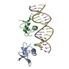
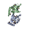
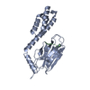
| Title | Crystal structure of Ruminococcus flavefaciens' type III complex containing the fifth cohesin from scaffoldin B and the dockerin from scaffoldin A | |||||||||
 Components Components | (Putative cellulosomal scaffoldin protein) x 2 | |||||||||
 Keywords Keywords |  PROTEIN BINDING / cellulosome / cohesin / dockerin / type III cohesin-dockerin / Coh-Doc / protein-protein interaction / cell adhesion PROTEIN BINDING / cellulosome / cohesin / dockerin / type III cohesin-dockerin / Coh-Doc / protein-protein interaction / cell adhesion | |||||||||
| Function / homology |  Function and homology information Function and homology informationpolysaccharide catabolic process / carbohydrate binding / extracellular regionSimilarity search - Function | |||||||||
| Biological species |  Ruminococcus flavefaciens FD-1 (bacteria) Ruminococcus flavefaciens FD-1 (bacteria) | |||||||||
| Method | X-RAY DIFFRACTION / SYNCHROTRON / MOLECULAR REPLACEMENT / molecular replacement / Resolution: 1.98 Å | |||||||||
 Authors Authors | Bule, P. / Carvalho, A.L. / Najmudin, S. / Fontes, C.M.G.A. | |||||||||
| Funding support |  Portugal, 2items Portugal, 2items
| |||||||||
 Citation Citation | Journal: Sci Rep / Year: 2018 Title: Higher order scaffoldin assembly in Ruminococcus flavefaciens cellulosome is coordinated by a discrete cohesin-dockerin interaction. Authors: Bule, P. / Pires, V.M.R. / Alves, V.D. / Carvalho, A.L. / Prates, J.A.M. / Ferreira, L.M.A. / Smith, S.P. / Gilbert, H.J. / Noach, I. / Bayer, E.A. / Najmudin, S. / Fontes, C.M.G.A. | |||||||||
| History |
|
- Structure visualization
| Structure viewer | Molecule: MolmilJmol/JSmol |
|---|
- Downloads & links
-Download
| PDBx/mmCIF format | 5n5p.cif.gz | 181.8 KB | Display | PDBx/mmCIF format |
|---|---|---|---|---|
| PDB format | pdb5n5p.ent.gz | 145.1 KB | Display | PDB format |
| PDBx/mmJSON format | 5n5p.json.gz | Tree view | PDBx/mmJSON format | |
| Others |  Other downloads Other downloads |
-Validation report
| Arichive directory | https://data.pdbj.org/pub/pdb/validation_reports/n5/5n5pftp://data.pdbj.org/pub/pdb/validation_reports/n5/5n5p | HTTPS FTP |
|---|
-Related structure data
| Related structure data |  5lxvS S: Starting model for refinement |
|---|---|
| Similar structure data |


-Links
 PDBj
PDBj
- Assembly
| Deposited unit | 
| ||||||||||||||||||||||||||||||||||||||||||||||||||||||||||
|---|---|---|---|---|---|---|---|---|---|---|---|---|---|---|---|---|---|---|---|---|---|---|---|---|---|---|---|---|---|---|---|---|---|---|---|---|---|---|---|---|---|---|---|---|---|---|---|---|---|---|---|---|---|---|---|---|---|---|---|
| 1 | 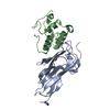
| ||||||||||||||||||||||||||||||||||||||||||||||||||||||||||
| 2 | 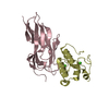
| ||||||||||||||||||||||||||||||||||||||||||||||||||||||||||
| Unit cell |
| ||||||||||||||||||||||||||||||||||||||||||||||||||||||||||
| Noncrystallographic symmetry (NCS) | NCS domain:
NCS domain segments: Component-ID: 0 / End auth comp-ID: GLY / End label comp-ID: GLY / Refine code: 0
NCS ensembles :
|
-Components
| #1: Protein | Mass: 14252.957 Da / Num. of mol.: 2 Fragment: ScaB Type III cohesin domain, UNP residues 739-876 Source method: isolated from a genetically manipulated source Source: (gene. exp.) Ruminococcus flavefaciens FD-1 (bacteria)Gene: scaB / Plasmid: pET28a / Production host: Escherichia coli BL21(DE3) (bacteria) / References: UniProt: A0AEF4#2: Protein | Mass: 9220.265 Da / Num. of mol.: 2 Fragment: ScaA Type III dockerin domain, UNP residues 648-730 Source method: isolated from a genetically manipulated source Source: (gene. exp.) Ruminococcus flavefaciens FD-1 (bacteria)Gene: scaA / Plasmid: pET28a / Production host: Escherichia coli BL21(DE3) (bacteria) / References: UniProt: A0AEF3#3: Chemical | ChemComp-CCN / | Acetonitrile  Mass: 41.052 Da / Num. of mol.: 1 / Source method: obtained synthetically / Formula: C2H3N Mass: 41.052 Da / Num. of mol.: 1 / Source method: obtained synthetically / Formula: C2H3N#4: Chemical | ChemComp-CA /   Mass: 40.078 Da / Num. of mol.: 4 / Source method: obtained synthetically / Formula: Ca Mass: 40.078 Da / Num. of mol.: 4 / Source method: obtained synthetically / Formula: Ca#5: Water | ChemComp-HOH / | Water Mass: 18.015 Da / Num. of mol.: 225 / Source method: isolated from a natural source / Formula: H2O Mass: 18.015 Da / Num. of mol.: 225 / Source method: isolated from a natural source / Formula: H2O |
|---|
-Experimental details
-Experiment
| Experiment | Method: X-RAY DIFFRACTION / Number of used crystals: 1 |
|---|
- Sample preparation
| Crystal | Density Matthews: 2.13 Å3/Da / Density % sol: 42.35 % / Mosaicity: 0.31 ° |
|---|---|
| Crystal grow | Temperature: 292 K / Method: vapor diffusion, hanging drop Details: 0.1M HEPES 7.5, 1.2M Sodium Citrate, 4% v/v Acetonitrile |
-Data collection
| Diffraction | Mean temperature: 100 K | |||||||||||||||||||||
|---|---|---|---|---|---|---|---|---|---|---|---|---|---|---|---|---|---|---|---|---|---|---|
| Diffraction source | Source: SYNCHROTRON / Site: SOLEIL  / Beamline: PROXIMA 1 / Wavelength: 0.82655 Å / Beamline: PROXIMA 1 / Wavelength: 0.82655 Å | |||||||||||||||||||||
| Detector | Type: DECTRIS PILATUS 6M / Detector: PIXEL / Date: Nov 15, 2014 | |||||||||||||||||||||
| Radiation | Protocol: SINGLE WAVELENGTH / Monochromatic (M) / Laue (L): M / Scattering type: x-ray | |||||||||||||||||||||
| Radiation wavelength | Wavelength: 0.82655 Å / Relative weight: 1 | |||||||||||||||||||||
| Reflection twin |
| |||||||||||||||||||||
| Reflection | Resolution: 1.4→47.63 Å / Num. obs: 26512 / % possible obs: 100 % / Redundancy: 6.9 % / CC1/2: 0.995 / Rmerge(I) obs: 0.098 / Rpim(I) all: 0.04 / Rrim(I) all: 0.106 / Net I/σ(I): 8.8 | |||||||||||||||||||||
| Reflection shell | Diffraction-ID: 1
|
-Phasing
| Phasing | Method: molecular replacement |
|---|
- Processing
| Software |
| |||||||||||||||||||||||||||||||||||||||||||||||||||||||||||||||||||||||||||||||||||||||||||||||||||||||||||||||||||||||||||||
|---|---|---|---|---|---|---|---|---|---|---|---|---|---|---|---|---|---|---|---|---|---|---|---|---|---|---|---|---|---|---|---|---|---|---|---|---|---|---|---|---|---|---|---|---|---|---|---|---|---|---|---|---|---|---|---|---|---|---|---|---|---|---|---|---|---|---|---|---|---|---|---|---|---|---|---|---|---|---|---|---|---|---|---|---|---|---|---|---|---|---|---|---|---|---|---|---|---|---|---|---|---|---|---|---|---|---|---|---|---|---|---|---|---|---|---|---|---|---|---|---|---|---|---|---|---|---|
| Refinement | Method to determine structure: MOLECULAR REPLACEMENT Starting model: 5lxv Resolution: 1.98→46.58 Å / Cor.coef. Fo:Fc: 0.956 / Cor.coef. Fo:Fc free: 0.934 / SU B: 9.114 / SU ML: 0.136 / Cross valid method: THROUGHOUT / σ(F): 0 / ESU R: 0.044 / ESU R Free: 0.035 Details: HYDROGENS HAVE BEEN ADDED IN THE RIDING POSITIONS U VALUES : WITH TLS ADDED
| |||||||||||||||||||||||||||||||||||||||||||||||||||||||||||||||||||||||||||||||||||||||||||||||||||||||||||||||||||||||||||||
| Solvent computation | Ion probe radii: 0.7 Å / Shrinkage radii: 0.7 Å / VDW probe radii: 1.1 Å | |||||||||||||||||||||||||||||||||||||||||||||||||||||||||||||||||||||||||||||||||||||||||||||||||||||||||||||||||||||||||||||
| Displacement parameters | Biso max: 114.41 Å2 / Biso mean: 33.69 Å2 / Biso min: 17.41 Å2
| |||||||||||||||||||||||||||||||||||||||||||||||||||||||||||||||||||||||||||||||||||||||||||||||||||||||||||||||||||||||||||||
| Refinement step | Cycle: final / Resolution: 1.98→46.58 Å
| |||||||||||||||||||||||||||||||||||||||||||||||||||||||||||||||||||||||||||||||||||||||||||||||||||||||||||||||||||||||||||||
| Refine LS restraints |
| |||||||||||||||||||||||||||||||||||||||||||||||||||||||||||||||||||||||||||||||||||||||||||||||||||||||||||||||||||||||||||||
| Refine LS restraints NCS | Refine-ID: X-RAY DIFFRACTION / Type: interatomic distance / Weight position: 0.05
| |||||||||||||||||||||||||||||||||||||||||||||||||||||||||||||||||||||||||||||||||||||||||||||||||||||||||||||||||||||||||||||
| LS refinement shell | Resolution: 1.976→2.027 Å / Rfactor Rfree error: 0 / Total num. of bins used: 20
| |||||||||||||||||||||||||||||||||||||||||||||||||||||||||||||||||||||||||||||||||||||||||||||||||||||||||||||||||||||||||||||
| Refinement TLS params. | Method: refined / Refine-ID: X-RAY DIFFRACTION
| |||||||||||||||||||||||||||||||||||||||||||||||||||||||||||||||||||||||||||||||||||||||||||||||||||||||||||||||||||||||||||||
| Refinement TLS group |
|
