 Movie
Movie Controller

[English] 日本語
 Yorodumi
- PDB-5lxv: Crystal structure of Ruminococcus flavefaciens scaffoldin C cohes... -
+ Open data

- Basic information
| Entry | Database: PDB / ID: 5lxv | |||||||||
|---|---|---|---|---|---|---|---|---|---|---|
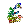
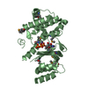
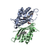
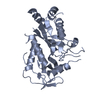
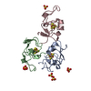
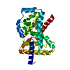
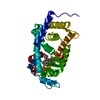
| Title | Crystal structure of Ruminococcus flavefaciens scaffoldin C cohesin in complex with a dockerin from an uncharacterized CBM-containing protein | |||||||||
 Components Components |
| |||||||||
 Keywords Keywords |  PROTEIN BINDING / cellulosome / cohesin / dockerin / type I cohesin-dockerin / Coh-Doc / protein-protein interaction PROTEIN BINDING / cellulosome / cohesin / dockerin / type I cohesin-dockerin / Coh-Doc / protein-protein interaction | |||||||||
| Function / homology |  Function and homology information Function and homology informationpolysaccharide catabolic process / hydrolase activity, hydrolyzing O-glycosyl compounds / carbohydrate binding / metal ion bindingSimilarity search - Function | |||||||||
| Biological species |  Ruminococcus flavefaciens FD-1 (bacteria) Ruminococcus flavefaciens FD-1 (bacteria) | |||||||||
| Method | X-RAY DIFFRACTION / SYNCHROTRON / MOLECULAR REPLACEMENT / molecular replacement / Resolution: 2.4 Å | |||||||||
 Authors Authors | Najmudin, S. / Bule, P. / Fontes, C.M.G.A. | |||||||||
| Funding support |  Portugal, 2items Portugal, 2items
| |||||||||
 Citation Citation | Journal: J. Biol. Chem. / Year: 2016 Title: Single Binding Mode Integration of Hemicellulose-degrading Enzymes via Adaptor Scaffoldins in Ruminococcus flavefaciens Cellulosome. Authors: Bule, P. / Alves, V.D. / Leitao, A. / Ferreira, L.M. / Bayer, E.A. / Smith, S.P. / Gilbert, H.J. / Najmudin, S. / Fontes, C.M. #1: Journal: Acta Crystallogr F Struct Biol Commun / Year: 2014 Title: Overexpression, crystallization and preliminary X-ray characterization of Ruminococcus flavefaciens scaffoldin C cohesin in complex with a dockerin from an uncharacterized CBM-containing protein. Authors: Bule, P. / Ruimy-Israeli, V. / Cardoso, V. / Bayer, E.A. / Fontes, C.M. / Najmudin, S. | |||||||||
| History |
|
- Structure visualization
| Structure viewer | Molecule: MolmilJmol/JSmol |
|---|
- Downloads & links
-Download
| PDBx/mmCIF format | 5lxv.cif.gz | 194.3 KB | Display | PDBx/mmCIF format |
|---|---|---|---|---|
| PDB format | pdb5lxv.ent.gz | 155.4 KB | Display | PDB format |
| PDBx/mmJSON format | 5lxv.json.gz | Tree view | PDBx/mmJSON format | |
| Others |  Other downloads Other downloads |
-Validation report
| Arichive directory | https://data.pdbj.org/pub/pdb/validation_reports/lx/5lxvftp://data.pdbj.org/pub/pdb/validation_reports/lx/5lxv | HTTPS FTP |
|---|
-Related structure data
| Related structure data | 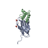 2cclS S: Starting model for refinement |
|---|---|
| Similar structure data |


-Links
 PDBj
PDBj
- Assembly
| Deposited unit | 
| ||||||||||||||||||||||||||||||||||||||||||||||||||||||||||||||||||||
|---|---|---|---|---|---|---|---|---|---|---|---|---|---|---|---|---|---|---|---|---|---|---|---|---|---|---|---|---|---|---|---|---|---|---|---|---|---|---|---|---|---|---|---|---|---|---|---|---|---|---|---|---|---|---|---|---|---|---|---|---|---|---|---|---|---|---|---|---|---|
| 1 | 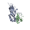
| ||||||||||||||||||||||||||||||||||||||||||||||||||||||||||||||||||||
| 2 | 
| ||||||||||||||||||||||||||||||||||||||||||||||||||||||||||||||||||||
| Unit cell |
| ||||||||||||||||||||||||||||||||||||||||||||||||||||||||||||||||||||
| Noncrystallographic symmetry (NCS) | NCS domain:
NCS domain segments: Component-ID: 0 / Refine code: 0
NCS ensembles :
|
-Components
| #1: Protein | Mass: 18571.367 Da / Num. of mol.: 2 / Fragment: ScaC Type I cohesin domain, UNP residues 29-204 Source method: isolated from a genetically manipulated source Source: (gene. exp.) Ruminococcus flavefaciens FD-1 (bacteria)Gene: scaC / Production host: Escherichia coli BL21(DE3) (bacteria) / References: UniProt: G9FCX2#2: Protein | Mass: 9791.689 Da / Num. of mol.: 2 / Fragment: Doc3: Type I dockerin domain Source method: isolated from a genetically manipulated source Source: (gene. exp.) Ruminococcus flavefaciens FD-1 (bacteria)Production host: Escherichia coli BL21(DE3) (bacteria) / References: UniProt: A0A1L1QK37*PLUS#3: Chemical | ChemComp-CA /   Mass: 40.078 Da / Num. of mol.: 4 / Source method: obtained synthetically / Formula: Ca Mass: 40.078 Da / Num. of mol.: 4 / Source method: obtained synthetically / Formula: Ca#4: Water | ChemComp-HOH / | Water Mass: 18.015 Da / Num. of mol.: 112 / Source method: isolated from a natural source / Formula: H2O Mass: 18.015 Da / Num. of mol.: 112 / Source method: isolated from a natural source / Formula: H2O |
|---|
-Experimental details
-Experiment
| Experiment | Method: X-RAY DIFFRACTION / Number of used crystals: 1 |
|---|
- Sample preparation
| Crystal | Density Matthews: 1.92 Å3/Da / Density % sol: 36.1 % |
|---|---|
| Crystal grow | Temperature: 292 K / Method: vapor diffusion, hanging drop / Details: 0.1 M potassium thiocyanate, 30% w/v PEG 2000 MME |
-Data collection
| Diffraction | Mean temperature: 100 K | ||||||||||||||||||
|---|---|---|---|---|---|---|---|---|---|---|---|---|---|---|---|---|---|---|---|
| Diffraction source | Source: SYNCHROTRON / Site: Diamond  / Beamline: I04-1 / Wavelength: 0.92 Å / Beamline: I04-1 / Wavelength: 0.92 Å | ||||||||||||||||||
| Detector | Type: DECTRIS PILATUS 6M-F / Detector: PIXEL / Date: Feb 8, 2014 | ||||||||||||||||||
| Radiation | Protocol: SINGLE WAVELENGTH / Monochromatic (M) / Laue (L): M / Scattering type: x-ray | ||||||||||||||||||
| Radiation wavelength | Wavelength: 0.92 Å / Relative weight: 1 | ||||||||||||||||||
| Reflection | Resolution: 2.4→59.59 Å / Num. obs: 17195 / % possible obs: 97.6 % / Redundancy: 4 % / CC1/2: 0.985 / Rmerge(I) obs: 0.08 / Net I/σ(I): 51.7 | ||||||||||||||||||
| Reflection shell |
|
-Phasing
| Phasing | Method: molecular replacement | |||||||||
|---|---|---|---|---|---|---|---|---|---|---|
| Phasing MR | Model details: Phaser MODE: MR_AUTO
|
- Processing
| Software |
| |||||||||||||||||||||||||||||||||||||||||||||||||||||||||||||||||||||||||||||||||||||||||||||||||||||||||||||||||||||||||||||
|---|---|---|---|---|---|---|---|---|---|---|---|---|---|---|---|---|---|---|---|---|---|---|---|---|---|---|---|---|---|---|---|---|---|---|---|---|---|---|---|---|---|---|---|---|---|---|---|---|---|---|---|---|---|---|---|---|---|---|---|---|---|---|---|---|---|---|---|---|---|---|---|---|---|---|---|---|---|---|---|---|---|---|---|---|---|---|---|---|---|---|---|---|---|---|---|---|---|---|---|---|---|---|---|---|---|---|---|---|---|---|---|---|---|---|---|---|---|---|---|---|---|---|---|---|---|---|
| Refinement | Method to determine structure: MOLECULAR REPLACEMENT Starting model: 2CCL Resolution: 2.4→57 Å / Cor.coef. Fo:Fc: 0.931 / Cor.coef. Fo:Fc free: 0.903 / SU B: 19.881 / SU ML: 0.229 / Cross valid method: THROUGHOUT / σ(F): 0 / ESU R: 0.808 / ESU R Free: 0.304 / Stereochemistry target values: MAXIMUM LIKELIHOOD Details: HYDROGENS HAVE BEEN ADDED IN THE RIDING POSITIONS U VALUES : WITH TLS ADDED
| |||||||||||||||||||||||||||||||||||||||||||||||||||||||||||||||||||||||||||||||||||||||||||||||||||||||||||||||||||||||||||||
| Solvent computation | Ion probe radii: 0.8 Å / Shrinkage radii: 0.8 Å / VDW probe radii: 1.1 Å / Solvent model: MASK | |||||||||||||||||||||||||||||||||||||||||||||||||||||||||||||||||||||||||||||||||||||||||||||||||||||||||||||||||||||||||||||
| Displacement parameters | Biso max: 90.22 Å2 / Biso mean: 35.648 Å2 / Biso min: 14.77 Å2
| |||||||||||||||||||||||||||||||||||||||||||||||||||||||||||||||||||||||||||||||||||||||||||||||||||||||||||||||||||||||||||||
| Refinement step | Cycle: final / Resolution: 2.4→57 Å
| |||||||||||||||||||||||||||||||||||||||||||||||||||||||||||||||||||||||||||||||||||||||||||||||||||||||||||||||||||||||||||||
| Refine LS restraints |
| |||||||||||||||||||||||||||||||||||||||||||||||||||||||||||||||||||||||||||||||||||||||||||||||||||||||||||||||||||||||||||||
| Refine LS restraints NCS | Refine-ID: X-RAY DIFFRACTION / Type: interatomic distance / Weight position: 0.05
| |||||||||||||||||||||||||||||||||||||||||||||||||||||||||||||||||||||||||||||||||||||||||||||||||||||||||||||||||||||||||||||
| LS refinement shell | Resolution: 2.4→2.462 Å / Total num. of bins used: 20
| |||||||||||||||||||||||||||||||||||||||||||||||||||||||||||||||||||||||||||||||||||||||||||||||||||||||||||||||||||||||||||||
| Refinement TLS params. | Method: refined / Refine-ID: X-RAY DIFFRACTION
| |||||||||||||||||||||||||||||||||||||||||||||||||||||||||||||||||||||||||||||||||||||||||||||||||||||||||||||||||||||||||||||
| Refinement TLS group |
|
