 Movie
Movie Controller

[English] 日本語
 Yorodumi
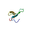
- PDB-5vti: Structure of Pin1 WW Domain Sequence 3 with [R,R]-ACPC Loop Subst... -
+ Open data

- Basic information
| Entry | Database: PDB / ID: 5vti | ||||||
|---|---|---|---|---|---|---|---|
| Title | Structure of Pin1 WW Domain Sequence 3 with [R,R]-ACPC Loop Substitution | ||||||
 Components Components | Peptidyl-prolyl cis-trans isomerase NIMA-interacting 1 | ||||||
 Keywords Keywords |  PROTEIN BINDING / beta amino acid / WW domain / phosphopeptide binding PROTEIN BINDING / beta amino acid / WW domain / phosphopeptide binding | ||||||
| Function / homology |  Function and homology informationcis-trans isomerase activity / phosphothreonine residue binding / negative regulation of cell motility / ubiquitin ligase activator activity / regulation of protein localization to nucleus / GTPase activating protein binding / postsynaptic cytosol / mitogen-activated protein kinase kinase binding / regulation of mitotic nuclear division / negative regulation of SMAD protein signal transduction ...cis-trans isomerase activity / phosphothreonine residue binding / negative regulation of cell motility / ubiquitin ligase activator activity / regulation of protein localization to nucleus / GTPase activating protein binding / postsynaptic cytosol / mitogen-activated protein kinase kinase binding / regulation of mitotic nuclear division / negative regulation of SMAD protein signal transduction / PI5P Regulates TP53 Acetylation / negative regulation of amyloid-beta formation / cytoskeletal motor activity / RHO GTPases Activate NADPH Oxidases / phosphoserine residue binding / protein peptidyl-prolyl isomerization / positive regulation of protein dephosphorylation / ciliary basal body / regulation of cytokinesis / negative regulation of protein binding / peptidylprolyl isomerase / peptidyl-prolyl cis-trans isomerase activity / Negative regulators of DDX58/IFIH1 signaling / phosphoprotein binding / synapse organization / regulation of protein phosphorylation / negative regulation of transforming growth factor beta receptor signaling pathway / regulation of protein stability / tau protein binding / neuron differentiation / negative regulation of protein catabolic process / negative regulation of ERK1 and ERK2 cascade / ISG15 antiviral mechanism / beta-catenin binding / positive regulation of GTPase activity / positive regulation of canonical Wnt signaling pathway / positive regulation of protein binding / midbody / regulation of gene expression / Regulation of TP53 Activity through Phosphorylation / protein stabilization / response to hypoxia / nuclear speck / positive regulation of protein phosphorylation / cell cycle / glutamatergic synapse / positive regulation of transcription by RNA polymerase II / nucleoplasm / nucleus / cytosol / cytoplasm Function and homology informationcis-trans isomerase activity / phosphothreonine residue binding / negative regulation of cell motility / ubiquitin ligase activator activity / regulation of protein localization to nucleus / GTPase activating protein binding / postsynaptic cytosol / mitogen-activated protein kinase kinase binding / regulation of mitotic nuclear division / negative regulation of SMAD protein signal transduction ...cis-trans isomerase activity / phosphothreonine residue binding / negative regulation of cell motility / ubiquitin ligase activator activity / regulation of protein localization to nucleus / GTPase activating protein binding / postsynaptic cytosol / mitogen-activated protein kinase kinase binding / regulation of mitotic nuclear division / negative regulation of SMAD protein signal transduction / PI5P Regulates TP53 Acetylation / negative regulation of amyloid-beta formation / cytoskeletal motor activity / RHO GTPases Activate NADPH Oxidases / phosphoserine residue binding / protein peptidyl-prolyl isomerization / positive regulation of protein dephosphorylation / ciliary basal body / regulation of cytokinesis / negative regulation of protein binding / peptidylprolyl isomerase / peptidyl-prolyl cis-trans isomerase activity / Negative regulators of DDX58/IFIH1 signaling / phosphoprotein binding / synapse organization / regulation of protein phosphorylation / negative regulation of transforming growth factor beta receptor signaling pathway / regulation of protein stability / tau protein binding / neuron differentiation / negative regulation of protein catabolic process / negative regulation of ERK1 and ERK2 cascade / ISG15 antiviral mechanism / beta-catenin binding / positive regulation of GTPase activity / positive regulation of canonical Wnt signaling pathway / positive regulation of protein binding / midbody / regulation of gene expression / Regulation of TP53 Activity through Phosphorylation / protein stabilization / response to hypoxia / nuclear speck / positive regulation of protein phosphorylation / cell cycle / glutamatergic synapse / positive regulation of transcription by RNA polymerase II / nucleoplasm / nucleus / cytosol / cytoplasmSimilarity search - Function | ||||||
| Biological species |  Homo sapiens (human) Homo sapiens (human) | ||||||
| Method | X-RAY DIFFRACTION / SYNCHROTRON / MOLECULAR REPLACEMENT / Resolution: 1.8 Å | ||||||
 Authors Authors | Mortenson, D.E. / Kreitler, D.F. / Thomas, N.C. / Gellman, S.H. / Forest, K.T. | ||||||
 Citation Citation | Journal: Chembiochem / Year: 2018 Title: Evaluation of beta-Amino Acid Replacements in Protein Loops: Effects on Conformational Stability and Structure. Authors: Mortenson, D.E. / Kreitler, D.F. / Thomas, N.C. / Guzei, I.A. / Gellman, S.H. / Forest, K.T. | ||||||
| History |
|
- Structure visualization
| Structure viewer | Molecule: MolmilJmol/JSmol |
|---|
- Downloads & links
-Download
| PDBx/mmCIF format | 5vti.cif.gz | 30.6 KB | Display | PDBx/mmCIF format |
|---|---|---|---|---|
| PDB format | pdb5vti.ent.gz | 18.8 KB | Display | PDB format |
| PDBx/mmJSON format | 5vti.json.gz | Tree view | PDBx/mmJSON format | |
| Others |  Other downloads Other downloads |
-Validation report
| Arichive directory | https://data.pdbj.org/pub/pdb/validation_reports/vt/5vtiftp://data.pdbj.org/pub/pdb/validation_reports/vt/5vti | HTTPS FTP |
|---|
-Related structure data
| Related structure data |  5vtjC  5vtkC  4gwtS C: citing same article ( S: Starting model for refinement |
|---|---|
| Similar structure data |


-Links
 PDBj
PDBj


- Assembly
| Deposited unit | 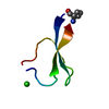
| ||||||||
|---|---|---|---|---|---|---|---|---|---|
| 1 |
| ||||||||
| Unit cell |
|
-Components
| #1: Protein/peptide | Mass: 3810.305 Da / Num. of mol.: 1 / Fragment: WW domain sequence 3 (UNP residues 6-39) / Source method: obtained synthetically / Source: (synth.) Homo sapiens (human) / References: UniProt: Q13526 |
|---|---|
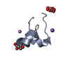
| #2: Chemical | ChemComp-CL / Chloride  Mass: 35.453 Da / Num. of mol.: 1 / Source method: obtained synthetically / Formula: Cl Mass: 35.453 Da / Num. of mol.: 1 / Source method: obtained synthetically / Formula: Cl |
| #3: Water | ChemComp-HOH / Water Mass: 18.015 Da / Num. of mol.: 42 / Source method: isolated from a natural source / Formula: H2O Mass: 18.015 Da / Num. of mol.: 42 / Source method: isolated from a natural source / Formula: H2O |
-Experimental details
-Experiment
| Experiment | Method: X-RAY DIFFRACTION / Number of used crystals: 1 |
|---|
- Sample preparation
| Crystal | Density Matthews: 2.83 Å3/Da / Density % sol: 56.58 % |
|---|---|
| Crystal grow | Temperature: 298 K / Method: vapor diffusion, hanging drop Details: 0.1 M Tris, pH 8.5, 3.0 M sodium chloride (Hampton Index #12), cryoprotection: dragged through Paratone-N prior to freezing |
-Data collection
| Diffraction | Mean temperature: 100 K |
|---|---|
| Diffraction source | Source: SYNCHROTRON / Site: APS  / Beamline: 21-ID-F / Wavelength: 0.9787 Å / Beamline: 21-ID-F / Wavelength: 0.9787 Å |
| Detector | Type: MARMOSAIC 225 mm CCD / Detector: CCD / Date: Jun 8, 2014 |
| Radiation | Monochromator: diamond(111) / Protocol: SINGLE WAVELENGTH / Monochromatic (M) / Laue (L): M / Scattering type: x-ray |
| Radiation wavelength | Wavelength: 0.9787 Å / Relative weight: 1 |
| Reflection | Resolution: 1.8→48.17 Å / Num. obs: 4398 / % possible obs: 99.9 % / Redundancy: 26.4 % / Biso Wilson estimate: 31.6 Å2 / Rpim(I) all: 0.015 / Rsym value: 0.076 / Net I/σ(I): 25.1 |
| Reflection shell | Resolution: 1.8→1.9 Å / Redundancy: 28.1 % / Mean I/σ(I) obs: 2.3 / Num. unique obs: 617 / Rpim(I) all: 0.36 / Rsym value: 1.902 / % possible all: 100 |
- Processing
| Software |
| ||||||||||||||||||||||||||||||||||||||||||||||||||||||||||||||||||||||||||||||||||||||||||||||||||||||||||||||||||||||||||||||||||||||||||||||||||||||||||||||||||||||||||||||||||||||
|---|---|---|---|---|---|---|---|---|---|---|---|---|---|---|---|---|---|---|---|---|---|---|---|---|---|---|---|---|---|---|---|---|---|---|---|---|---|---|---|---|---|---|---|---|---|---|---|---|---|---|---|---|---|---|---|---|---|---|---|---|---|---|---|---|---|---|---|---|---|---|---|---|---|---|---|---|---|---|---|---|---|---|---|---|---|---|---|---|---|---|---|---|---|---|---|---|---|---|---|---|---|---|---|---|---|---|---|---|---|---|---|---|---|---|---|---|---|---|---|---|---|---|---|---|---|---|---|---|---|---|---|---|---|---|---|---|---|---|---|---|---|---|---|---|---|---|---|---|---|---|---|---|---|---|---|---|---|---|---|---|---|---|---|---|---|---|---|---|---|---|---|---|---|---|---|---|---|---|---|---|---|---|---|
| Refinement | Method to determine structure: MOLECULAR REPLACEMENT Starting model: PDB entry 4GWT Resolution: 1.8→34.06 Å / Cor.coef. Fo:Fc: 0.948 / Cor.coef. Fo:Fc free: 0.929 / SU B: 7.466 / SU ML: 0.119 / Cross valid method: THROUGHOUT / ESU R: 0.152 / ESU R Free: 0.148 / Details: HYDROGENS HAVE BEEN ADDED IN THE RIDING POSITIONS
| ||||||||||||||||||||||||||||||||||||||||||||||||||||||||||||||||||||||||||||||||||||||||||||||||||||||||||||||||||||||||||||||||||||||||||||||||||||||||||||||||||||||||||||||||||||||
| Solvent computation | Ion probe radii: 0.8 Å / Shrinkage radii: 0.8 Å / VDW probe radii: 1.2 Å | ||||||||||||||||||||||||||||||||||||||||||||||||||||||||||||||||||||||||||||||||||||||||||||||||||||||||||||||||||||||||||||||||||||||||||||||||||||||||||||||||||||||||||||||||||||||
| Displacement parameters | Biso mean: 43.65 Å2
| ||||||||||||||||||||||||||||||||||||||||||||||||||||||||||||||||||||||||||||||||||||||||||||||||||||||||||||||||||||||||||||||||||||||||||||||||||||||||||||||||||||||||||||||||||||||
| Refinement step | Cycle: 1 / Resolution: 1.8→34.06 Å
| ||||||||||||||||||||||||||||||||||||||||||||||||||||||||||||||||||||||||||||||||||||||||||||||||||||||||||||||||||||||||||||||||||||||||||||||||||||||||||||||||||||||||||||||||||||||
| Refine LS restraints |
|