 Movie
Movie Controller

[English] 日本語
 Yorodumi
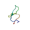
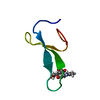
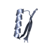
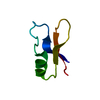
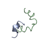
- PDB-4gwt: Structure of racemic Pin1 WW domain cocrystallized with DL-malic acid -
+ Open data

- Basic information
| Entry | Database: PDB / ID: 4gwt | ||||||
|---|---|---|---|---|---|---|---|
| Title | Structure of racemic Pin1 WW domain cocrystallized with DL-malic acid | ||||||
 Components Components | Peptidyl-prolyl cis-trans isomerase NIMA-interacting 1 | ||||||
 Keywords Keywords |  PROTEIN BINDING / racemic crystallization / WW domain / proline phosphoSer/Thr binding PROTEIN BINDING / racemic crystallization / WW domain / proline phosphoSer/Thr binding | ||||||
| Function / homology |  Function and homology informationcis-trans isomerase activity / phosphothreonine residue binding / negative regulation of cell motility / ubiquitin ligase activator activity / regulation of protein localization to nucleus / GTPase activating protein binding / postsynaptic cytosol / mitogen-activated protein kinase kinase binding / regulation of mitotic nuclear division / negative regulation of SMAD protein signal transduction ...cis-trans isomerase activity / phosphothreonine residue binding / negative regulation of cell motility / ubiquitin ligase activator activity / regulation of protein localization to nucleus / GTPase activating protein binding / postsynaptic cytosol / mitogen-activated protein kinase kinase binding / regulation of mitotic nuclear division / negative regulation of SMAD protein signal transduction / PI5P Regulates TP53 Acetylation / negative regulation of amyloid-beta formation / cytoskeletal motor activity / RHO GTPases Activate NADPH Oxidases / phosphoserine residue binding / protein peptidyl-prolyl isomerization / positive regulation of protein dephosphorylation / ciliary basal body / regulation of cytokinesis / negative regulation of protein binding / peptidylprolyl isomerase / peptidyl-prolyl cis-trans isomerase activity / Negative regulators of DDX58/IFIH1 signaling / phosphoprotein binding / synapse organization / regulation of protein phosphorylation / negative regulation of transforming growth factor beta receptor signaling pathway / regulation of protein stability / tau protein binding / neuron differentiation / negative regulation of protein catabolic process / negative regulation of ERK1 and ERK2 cascade / ISG15 antiviral mechanism / beta-catenin binding / positive regulation of GTPase activity / positive regulation of canonical Wnt signaling pathway / positive regulation of protein binding / midbody / regulation of gene expression / Regulation of TP53 Activity through Phosphorylation / protein stabilization / response to hypoxia / nuclear speck / positive regulation of protein phosphorylation / cell cycle / glutamatergic synapse / positive regulation of transcription by RNA polymerase II / nucleoplasm / nucleus / cytosol / cytoplasm Function and homology informationcis-trans isomerase activity / phosphothreonine residue binding / negative regulation of cell motility / ubiquitin ligase activator activity / regulation of protein localization to nucleus / GTPase activating protein binding / postsynaptic cytosol / mitogen-activated protein kinase kinase binding / regulation of mitotic nuclear division / negative regulation of SMAD protein signal transduction ...cis-trans isomerase activity / phosphothreonine residue binding / negative regulation of cell motility / ubiquitin ligase activator activity / regulation of protein localization to nucleus / GTPase activating protein binding / postsynaptic cytosol / mitogen-activated protein kinase kinase binding / regulation of mitotic nuclear division / negative regulation of SMAD protein signal transduction / PI5P Regulates TP53 Acetylation / negative regulation of amyloid-beta formation / cytoskeletal motor activity / RHO GTPases Activate NADPH Oxidases / phosphoserine residue binding / protein peptidyl-prolyl isomerization / positive regulation of protein dephosphorylation / ciliary basal body / regulation of cytokinesis / negative regulation of protein binding / peptidylprolyl isomerase / peptidyl-prolyl cis-trans isomerase activity / Negative regulators of DDX58/IFIH1 signaling / phosphoprotein binding / synapse organization / regulation of protein phosphorylation / negative regulation of transforming growth factor beta receptor signaling pathway / regulation of protein stability / tau protein binding / neuron differentiation / negative regulation of protein catabolic process / negative regulation of ERK1 and ERK2 cascade / ISG15 antiviral mechanism / beta-catenin binding / positive regulation of GTPase activity / positive regulation of canonical Wnt signaling pathway / positive regulation of protein binding / midbody / regulation of gene expression / Regulation of TP53 Activity through Phosphorylation / protein stabilization / response to hypoxia / nuclear speck / positive regulation of protein phosphorylation / cell cycle / glutamatergic synapse / positive regulation of transcription by RNA polymerase II / nucleoplasm / nucleus / cytosol / cytoplasmSimilarity search - Function | ||||||
| Biological species |  Homo sapiens (human) Homo sapiens (human) | ||||||
| Method | X-RAY DIFFRACTION / MOLECULAR REPLACEMENT / Resolution: 2.25 Å | ||||||
 Authors Authors | Mortenson, D.E. / Yun, H.G. / Gellman, S.H. / Forest, K.T. | ||||||
 Citation Citation | Journal: Acta Crystallogr.,Sect.D / Year: 2013 Title: Evidence for small-molecule-mediated loop stabilization in the structure of the isolated Pin1 WW domain. Authors: Mortenson, D.E. / Kreitler, D.F. / Yun, H.G. / Gellman, S.H. / Forest, K.T. | ||||||
| History |
|
- Structure visualization
| Structure viewer | Molecule: MolmilJmol/JSmol |
|---|
- Downloads & links
-Download
| PDBx/mmCIF format | 4gwt.cif.gz | 19.8 KB | Display | PDBx/mmCIF format |
|---|---|---|---|---|
| PDB format | pdb4gwt.ent.gz | 11.1 KB | Display | PDB format |
| PDBx/mmJSON format | 4gwt.json.gz | Tree view | PDBx/mmJSON format | |
| Others |  Other downloads Other downloads |
-Validation report
| Arichive directory | https://data.pdbj.org/pub/pdb/validation_reports/gw/4gwtftp://data.pdbj.org/pub/pdb/validation_reports/gw/4gwt | HTTPS FTP |
|---|
-Related structure data
| Related structure data |  4gwvC 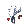 2idhS S: Starting model for refinement C: citing same article ( |
|---|---|
| Similar structure data |


-Links
 PDBj
PDBj


- Assembly
| Deposited unit | 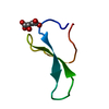
| ||||||||
|---|---|---|---|---|---|---|---|---|---|
| 1 |
| ||||||||
| Unit cell |
|
-Components
| #1: Protein/peptide | Mass: 4174.641 Da / Num. of mol.: 1 / Fragment: WW domain from Pin1, (6-39) / Source method: obtained synthetically / Details: Generated via solid-phase peptide synthesis / Source: (synth.) Homo sapiens (human) / References: UniProt: Q13526, peptidylprolyl isomerase |
|---|---|
| #2: Chemical | ChemComp-LMR / (Malic acid  Mass: 134.087 Da / Num. of mol.: 1 / Source method: obtained synthetically / Formula: C4H6O5 Mass: 134.087 Da / Num. of mol.: 1 / Source method: obtained synthetically / Formula: C4H6O5 |
| #3: Water | ChemComp-HOH / Water Mass: 18.015 Da / Num. of mol.: 11 / Source method: isolated from a natural source / Formula: H2O Mass: 18.015 Da / Num. of mol.: 11 / Source method: isolated from a natural source / Formula: H2O |
-Experimental details
-Experiment
| Experiment | Method: X-RAY DIFFRACTION / Number of used crystals: 1 |
|---|
- Sample preparation
| Crystal | Density Matthews: 2.52 Å3/Da / Density % sol: 51.14 % |
|---|---|
| Crystal grow | Temperature: 298 K / Method: vapor diffusion, hanging drop / pH: 7 Details: Peptide stock at 5 mg/mL (2.5 mg/mL L-peptide + 2.5 mg/L D-peptide), crystallized from 2.1 M DL-malic acid pH 7.0, VAPOR DIFFUSION, HANGING DROP, temperature 298K |
-Data collection
| Diffraction | Mean temperature: 298 K | |||||||||||||||||||||||||||||||||||||||||||||||||||||||||||||||||||||||||||||||||||||||||||||||||||||||||||||||||||||||||||||||||||||||||||||||||||
|---|---|---|---|---|---|---|---|---|---|---|---|---|---|---|---|---|---|---|---|---|---|---|---|---|---|---|---|---|---|---|---|---|---|---|---|---|---|---|---|---|---|---|---|---|---|---|---|---|---|---|---|---|---|---|---|---|---|---|---|---|---|---|---|---|---|---|---|---|---|---|---|---|---|---|---|---|---|---|---|---|---|---|---|---|---|---|---|---|---|---|---|---|---|---|---|---|---|---|---|---|---|---|---|---|---|---|---|---|---|---|---|---|---|---|---|---|---|---|---|---|---|---|---|---|---|---|---|---|---|---|---|---|---|---|---|---|---|---|---|---|---|---|---|---|---|---|---|---|
| Diffraction source | Source: ROTATING ANODE / Type: BRUKER AXS MICROSTAR / Wavelength: 1.5418 Å | |||||||||||||||||||||||||||||||||||||||||||||||||||||||||||||||||||||||||||||||||||||||||||||||||||||||||||||||||||||||||||||||||||||||||||||||||||
| Detector | Type: BRUKER SMART 6000 / Detector: CCD / Date: Mar 30, 2011 | |||||||||||||||||||||||||||||||||||||||||||||||||||||||||||||||||||||||||||||||||||||||||||||||||||||||||||||||||||||||||||||||||||||||||||||||||||
| Radiation | Monochromator: Bruker Microstar AXS optics / Protocol: SINGLE WAVELENGTH / Monochromatic (M) / Laue (L): M / Scattering type: x-ray | |||||||||||||||||||||||||||||||||||||||||||||||||||||||||||||||||||||||||||||||||||||||||||||||||||||||||||||||||||||||||||||||||||||||||||||||||||
| Radiation wavelength | Wavelength: 1.5418 Å / Relative weight: 1 | |||||||||||||||||||||||||||||||||||||||||||||||||||||||||||||||||||||||||||||||||||||||||||||||||||||||||||||||||||||||||||||||||||||||||||||||||||
| Reflection | Resolution: 2.25→33.32 Å / Num. all: 4031 / Num. obs: 4031 / % possible obs: 100 % / Observed criterion σ(F): 0 / Observed criterion σ(I): 0 / Redundancy: 13.3 % / Biso Wilson estimate: 41.6 Å2 / Rsym value: 0.069 / Χ2: 0.989 / Net I/σ(I): 22.85 | |||||||||||||||||||||||||||||||||||||||||||||||||||||||||||||||||||||||||||||||||||||||||||||||||||||||||||||||||||||||||||||||||||||||||||||||||||
| Reflection shell |
|
- Processing
| Software |
| |||||||||||||||||||||||||||||||||||||||||||||
|---|---|---|---|---|---|---|---|---|---|---|---|---|---|---|---|---|---|---|---|---|---|---|---|---|---|---|---|---|---|---|---|---|---|---|---|---|---|---|---|---|---|---|---|---|---|---|
| Refinement | Method to determine structure: MOLECULAR REPLACEMENT Starting model: PDB ENTRY 2IDH, CORE BETA-SHEET FRAGMENT (RESI 260-263,268-272,277-279) Resolution: 2.25→33.32 Å / Cor.coef. Fo:Fc: 0.955 / Cor.coef. Fo:Fc free: 0.947 / Occupancy max: 1 / Occupancy min: 1 / SU B: 3.765 / SU ML: 0.086 / Cross valid method: THROUGHOUT / σ(F): 0 / ESU R: 0.187 / ESU R Free: 0.168 / Stereochemistry target values: MAXIMUM LIKELIHOOD
| |||||||||||||||||||||||||||||||||||||||||||||
| Solvent computation | Ion probe radii: 0.8 Å / Shrinkage radii: 0.8 Å / VDW probe radii: 1.2 Å / Solvent model: MASK | |||||||||||||||||||||||||||||||||||||||||||||
| Displacement parameters | Biso max: 112.34 Å2 / Biso mean: 39.5828 Å2 / Biso min: 21.37 Å2
| |||||||||||||||||||||||||||||||||||||||||||||
| Refine analyze |
| |||||||||||||||||||||||||||||||||||||||||||||
| Refinement step | Cycle: LAST / Resolution: 2.25→33.32 Å
| |||||||||||||||||||||||||||||||||||||||||||||
| Refine LS restraints |
| |||||||||||||||||||||||||||||||||||||||||||||
| LS refinement shell | Resolution: 2.25→2.371 Å / Total num. of bins used: 10
|
