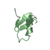
Deposited unit
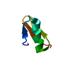
A: Amyloid beta A4 protein-binding family B member 1
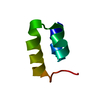
B: Amyloid beta A4 protein-binding family B member 1
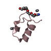
C: Amyloid beta A4 protein-binding family B member 1
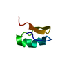
D: Amyloid beta A4 protein-binding family B member 1
E: Amyloid beta A4 protein-binding family B member 1
F: Amyloid beta A4 protein-binding family B member 1
G: Amyloid beta A4 protein-binding family B member 1
H: Amyloid beta A4 protein-binding family B member 1
hetero molecules Summary Component details
Theoretical mass Number of molelcules Total (without water) 35,108 17 Polymers 33,556 8 Non-polymers 1,552 9 Water 2,144 119
1
A: Amyloid beta A4 protein-binding family B member 1
hetero molecules Summary Component details Symmetry operations
Theoretical mass Number of molelcules Total (without water) 4,485 3 Polymers 4,195 1 Non-polymers 290 2 Water 18 1
Type Name Symmetry operation Number identity operation 1_555 x,y,z 1
2
B: Amyloid beta A4 protein-binding family B member 1 Summary Component details Symmetry operations
Theoretical mass Number of molelcules Total (without water) 4,195 1 Polymers 4,195 1 Non-polymers 0 0 Water 18 1
Type Name Symmetry operation Number identity operation 1_555 x,y,z 1
3
C: Amyloid beta A4 protein-binding family B member 1
hetero molecules Summary Component details Symmetry operations
Theoretical mass Number of molelcules Total (without water) 4,389 2 Polymers 4,195 1 Non-polymers 194 1 Water 18 1
Type Name Symmetry operation Number identity operation 1_555 x,y,z 1
4
D: Amyloid beta A4 protein-binding family B member 1
hetero molecules Summary Component details Symmetry operations
Theoretical mass Number of molelcules Total (without water) 4,485 3 Polymers 4,195 1 Non-polymers 290 2 Water 18 1
Type Name Symmetry operation Number identity operation 1_555 x,y,z 1
5
E: Amyloid beta A4 protein-binding family B member 1
hetero molecules Summary Component details Symmetry operations
Theoretical mass Number of molelcules Total (without water) 4,389 2 Polymers 4,195 1 Non-polymers 194 1 Water 18 1
Type Name Symmetry operation Number identity operation 1_555 x,y,z 1
6
F: Amyloid beta A4 protein-binding family B member 1
hetero molecules Summary Component details Symmetry operations
Theoretical mass Number of molelcules Total (without water) 4,389 2 Polymers 4,195 1 Non-polymers 194 1 Water 18 1
Type Name Symmetry operation Number identity operation 1_555 x,y,z 1
7
G: Amyloid beta A4 protein-binding family B member 1 Summary Component details Symmetry operations
Theoretical mass Number of molelcules Total (without water) 4,195 1 Polymers 4,195 1 Non-polymers 0 0 Water 18 1
Type Name Symmetry operation Number identity operation 1_555 x,y,z 1
8
H: Amyloid beta A4 protein-binding family B member 1
hetero molecules Summary Component details Symmetry operations
Theoretical mass Number of molelcules Total (without water) 4,583 3 Polymers 4,195 1 Non-polymers 388 2 Water 18 1
Type Name Symmetry operation Number identity operation 1_555 x,y,z 1
Unit cell Length a, b, c (Å) 75.610, 75.610, 226.489 Angle α, β, γ (deg.) 90.00, 90.00, 120.00 Int Tables number 182 Space group name H-M P63 22
Components on special symmetry positions ID Model Components 1 1 H -312-HOH
Noncrystallographic symmetry (NCS) NCS domain ID Ens-ID Details 1 1 A2 1 G3 1 F1 2 E2 2 H3 2 C1 3 A2 3 B1 4 C2 4 D
NCS domain segments Component-ID / Beg auth comp-ID / Beg label comp-ID / End auth comp-ID / End label comp-ID / Refine code / Auth seq-ID / Label seq-ID
Dom-ID Ens-ID Auth asym-ID Label asym-ID 1 1 AA2 1 GG3 1 FF1 2 EE2 2 HH3 2 CC1 3 AA2 3 BB1 4 CC2 4 DD
NCS ensembles
 Movie
Movie Controller


 Open data
 Basic information
 Components
 Keywords
 PROTEIN BINDING /
  Function and homology information
 Authors
 Citation
 Structure visualization
 Downloads & links
 Other downloads
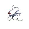

 PDBj
PDBj

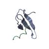
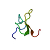
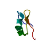
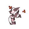
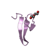
 Assembly
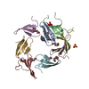


 Mass: 96.063 Da / Num. of mol.: 2 / Source method: obtained synthetically / Formula: SO4
 Mass: 194.226 Da / Num. of mol.: 7 / Source method: obtained synthetically / Formula: C8H18O5 / Comment: precipitant*YM
 Mass: 18.015 Da / Num. of mol.: 119 / Source method: isolated from a natural source / Formula: H2O
 Sample preparation
 Processing