 Movie
Movie Controller

[English] 日本語
 Yorodumi
- PDB-5vln: NMR structure of the N-domain of troponin C bound to switch regio... -
+ Open data

- Basic information
| Entry | Database: PDB / ID: 5vln | ||||||
|---|---|---|---|---|---|---|---|
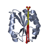
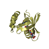
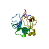
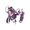
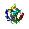
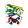
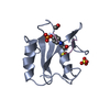
| Title | NMR structure of the N-domain of troponin C bound to switch region of troponin I | ||||||
 Components Components | Troponin C, slow skeletal and cardiac muscles,Troponin I, cardiac muscle | ||||||
 Keywords Keywords | METAL BINDING PROTEIN / cardiac troponin calcium binding protein EF hand / CELL INVASION | ||||||
| Function / homology |  Function and homology information Function and homology informationregulation of systemic arterial blood pressure by ischemic conditions / regulation of muscle filament sliding speed / troponin T binding / diaphragm contraction / troponin C binding / regulation of ATP-dependent activity / cardiac Troponin complex / cardiac myofibril / regulation of smooth muscle contraction / troponin complex ...regulation of systemic arterial blood pressure by ischemic conditions / regulation of muscle filament sliding speed / troponin T binding / diaphragm contraction / troponin C binding / regulation of ATP-dependent activity / cardiac Troponin complex / cardiac myofibril / regulation of smooth muscle contraction / troponin complex / regulation of muscle contraction / transition between fast and slow fiber / negative regulation of ATP-dependent activity / Striated Muscle Contraction / response to metal ion / regulation of cardiac muscle contraction by calcium ion signaling / myosin II complex / ventricular cardiac muscle tissue morphogenesis / heart contraction / troponin I binding / skeletal muscle contraction / calcium channel inhibitor activity / vasculogenesis / cardiac muscle contraction / Ion homeostasis / sarcomere / intracellular calcium ion homeostasis / calcium-dependent protein binding / actin filament binding / actin binding / heart development / protein domain specific binding / calcium ion binding / protein kinase binding / protein homodimerization activity / cytosolSimilarity search - Function | ||||||
| Biological species |  Homo sapiens (human) Homo sapiens (human) | ||||||
| Method | SOLUTION NMR / molecular dynamics | ||||||
 Authors Authors | Cai, F. / Hwang, P.M. / Sykes, B.D. | ||||||
| Funding support |  Canada, 1items Canada, 1items
| ||||||
 Citation Citation | Journal: J. Mol. Cell. Cardiol. / Year: 2016 Title: Structures reveal details of small molecule binding to cardiac troponin. Authors: Cai, F. / Li, M.X. / Pineda-Sanabria, S.E. / Gelozia, S. / Lindert, S. / West, F. / Sykes, B.D. / Hwang, P.M. | ||||||
| History |
|
- Structure visualization
| Structure viewer | Molecule: MolmilJmol/JSmol |
|---|
- Downloads & links
-Download
| PDBx/mmCIF format | 5vln.cif.gz | 298.4 KB | Display | PDBx/mmCIF format |
|---|---|---|---|---|
| PDB format | pdb5vln.ent.gz | 246.1 KB | Display | PDB format |
| PDBx/mmJSON format | 5vln.json.gz | Tree view | PDBx/mmJSON format | |
| Others |  Other downloads Other downloads |
-Validation report
| Arichive directory | https://data.pdbj.org/pub/pdb/validation_reports/vl/5vlnftp://data.pdbj.org/pub/pdb/validation_reports/vl/5vln | HTTPS FTP |
|---|
-Related structure data
| Related structure data |  5w88C  5wclC C: citing same article ( |
|---|---|
| Similar structure data | |
| Other databases |
|


-Links
 PDBj
PDBj


- Assembly
| Deposited unit | 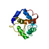
| |||||||||
|---|---|---|---|---|---|---|---|---|---|---|
| 1 |
| |||||||||
| NMR ensembles |
|
-Components
| #1: Protein | / TN-C / Cardiac troponin I Mass: 13495.394 Da / Num. of mol.: 1 / Mutation: C35S C84S,C35S C84S,C35S C84S,C35S C84S Source method: isolated from a genetically manipulated source Source: (gene. exp.) Homo sapiens (human) / Gene: TNNC1, TNNC, TNNI3, TNNC1 / Plasmid: pD444 / Production host:  Escherichia coli (E. coli) / Strain (production host): LB21(DE3), BLB21(DE3) / References: UniProt: P63316, UniProt: P19429 Escherichia coli (E. coli) / Strain (production host): LB21(DE3), BLB21(DE3) / References: UniProt: P63316, UniProt: P19429 |
|---|
-Experimental details
-Experiment
| Experiment | Method: SOLUTION NMR | ||||||||||||||||||||||||||||||||||||||||||||||||||||||||||||||||||||||||||||||||||||
|---|---|---|---|---|---|---|---|---|---|---|---|---|---|---|---|---|---|---|---|---|---|---|---|---|---|---|---|---|---|---|---|---|---|---|---|---|---|---|---|---|---|---|---|---|---|---|---|---|---|---|---|---|---|---|---|---|---|---|---|---|---|---|---|---|---|---|---|---|---|---|---|---|---|---|---|---|---|---|---|---|---|---|---|---|---|
| NMR experiment |
|
- Sample preparation
| Details |
| ||||||||||||||||||||||||||||||||||||||||||||||||||||||||||||||||||||||
|---|---|---|---|---|---|---|---|---|---|---|---|---|---|---|---|---|---|---|---|---|---|---|---|---|---|---|---|---|---|---|---|---|---|---|---|---|---|---|---|---|---|---|---|---|---|---|---|---|---|---|---|---|---|---|---|---|---|---|---|---|---|---|---|---|---|---|---|---|---|---|---|
| Sample |
|