 Movie
Movie Controller

[English] 日本語
 Yorodumi
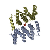
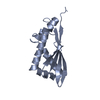
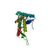
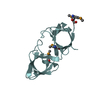
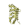
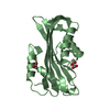
- PDB-5v7c: Crystal structure of LARP1-unique domain DM15 bound 5'TOP RNA sequence -
+ Open data

- Basic information
| Entry | Database: PDB / ID: 5v7c | ||||||
|---|---|---|---|---|---|---|---|
| Title | Crystal structure of LARP1-unique domain DM15 bound 5'TOP RNA sequence | ||||||
 Components Components |
| ||||||
 Keywords Keywords |  RNA BINDING PROTEIN / Cap-binding / RNA-binding / DM15 / 5'TOP RNA BINDING PROTEIN / Cap-binding / RNA-binding / DM15 / 5'TOP | ||||||
| Function / homology |  Function and homology information Function and homology informationcellular response to rapamycin / translation activator activity / eukaryotic initiation factor 4E binding / RNA cap binding / TORC1 signaling / response to amino acid starvation / RNA 7-methylguanosine cap binding / mRNA stabilization / post-transcriptional regulation of gene expression / positive regulation of macroautophagy ...cellular response to rapamycin / translation activator activity / eukaryotic initiation factor 4E binding / RNA cap binding / TORC1 signaling / response to amino acid starvation / RNA 7-methylguanosine cap binding / mRNA stabilization / post-transcriptional regulation of gene expression / positive regulation of macroautophagy / ribosomal small subunit binding / TOR signaling / positive regulation of translational initiation / positive regulation of viral genome replication / negative regulation of translational initiation / translational initiation / translation initiation factor binding / mRNA 3'-UTR binding / positive regulation of translation / mRNA 5'-UTR binding / cytoplasmic stress granule / cell population proliferation / negative regulation of translation / cadherin binding / SARS-CoV-2 activates/modulates innate and adaptive immune responses / RNA binding / membrane / cytosol / cytoplasmSimilarity search - Function | ||||||
| Biological species |  Homo sapiens (human) Homo sapiens (human) | ||||||
| Method | X-RAY DIFFRACTION / MOLECULAR REPLACEMENT / Resolution: 2.59 Å | ||||||
 Authors Authors | Berman, A.J. / Lahr, R.M. / Al-Ashtal, H.A. | ||||||
 Citation Citation | Journal: Elife / Year: 2017 Title: La-related protein 1 (LARP1) binds the mRNA cap, blocking eIF4F assembly on TOP mRNAs. Authors: Lahr, R.M. / Fonseca, B.D. / Ciotti, G.E. / Al-Ashtal, H.A. / Jia, J.J. / Niklaus, M.R. / Blagden, S.P. / Alain, T. / Berman, A.J. | ||||||
| History |
|
- Structure visualization
| Structure viewer | Molecule: MolmilJmol/JSmol |
|---|
- Downloads & links
-Download
| PDBx/mmCIF format | 5v7c.cif.gz | 50.4 KB | Display | PDBx/mmCIF format |
|---|---|---|---|---|
| PDB format | pdb5v7c.ent.gz | 32.6 KB | Display | PDB format |
| PDBx/mmJSON format | 5v7c.json.gz | Tree view | PDBx/mmJSON format | |
| Others |  Other downloads Other downloads |
-Validation report
| Arichive directory | https://data.pdbj.org/pub/pdb/validation_reports/v7/5v7cftp://data.pdbj.org/pub/pdb/validation_reports/v7/5v7c | HTTPS FTP |
|---|
-Related structure data
| Related structure data |  5v4rC 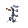 5v87C 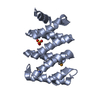 5c0vS C: citing same article ( S: Starting model for refinement |
|---|---|
| Similar structure data |


-Links
 PDBj
PDBj
- Assembly
| Deposited unit | 
| ||||||||
|---|---|---|---|---|---|---|---|---|---|
| 1 |
| ||||||||
| Unit cell |
|
-Components
| #1: Protein | Mass: 19616.281 Da / Num. of mol.: 1 Source method: isolated from a genetically manipulated source Details: DM15 / Source: (gene. exp.) Homo sapiens (human) / Gene: LARP1, KIAA0731, LARP / Production host:  Escherichia coli BL21(DE3) (bacteria) / References: UniProt: Q6PKG0 Escherichia coli BL21(DE3) (bacteria) / References: UniProt: Q6PKG0 |
|---|---|
| #2: RNA chain | Mass: 2440.457 Da / Num. of mol.: 1 / Source method: obtained synthetically / Source: (synth.) Homo sapiens (human) |
| #3: Water | ChemComp-HOH / Water Mass: 18.015 Da / Num. of mol.: 7 / Source method: isolated from a natural source / Formula: H2O Mass: 18.015 Da / Num. of mol.: 7 / Source method: isolated from a natural source / Formula: H2O |
-Experimental details
-Experiment
| Experiment | Method: X-RAY DIFFRACTION / Number of used crystals: 1 |
|---|
- Sample preparation
| Crystal | Density Matthews: 1.97 Å3/Da / Density % sol: 37.63 % |
|---|---|
| Crystal grow | Temperature: 293 K / Method: vapor diffusion, sitting drop / Details: 5 mM MgCl2, 50 mM HEPES, pH 7.0, 25 % PEG MME 550 |
-Data collection
| Diffraction | Mean temperature: 100 K |
|---|---|
| Diffraction source | Source: ROTATING ANODE / Type: RIGAKU FR-E DW / Wavelength: 1.54 Å |
| Detector | Type: RIGAKU SATURN 944 / Detector: CCD / Date: Jan 1, 2015 |
| Radiation | Protocol: SINGLE WAVELENGTH / Monochromatic (M) / Laue (L): M / Scattering type: x-ray |
| Radiation wavelength | Wavelength: 1.54 Å / Relative weight: 1 |
| Reflection | Resolution: 2.59→37.864 Å / Num. obs: 5272 / % possible obs: 98.5 % / Redundancy: 4.3 % / Net I/σ(I): 8.8 |
| Reflection shell | Highest resolution: 2.59 Å / Redundancy: 2.1 % / Mean I/σ(I) obs: 1.36 / % possible all: 87.1 |
- Processing
| Software |
| |||||||||||||||||||||||||||||||||||||||||||||||||
|---|---|---|---|---|---|---|---|---|---|---|---|---|---|---|---|---|---|---|---|---|---|---|---|---|---|---|---|---|---|---|---|---|---|---|---|---|---|---|---|---|---|---|---|---|---|---|---|---|---|---|
| Refinement | Method to determine structure: MOLECULAR REPLACEMENT Starting model: 5C0V Resolution: 2.59→37.864 Å / SU ML: 0.27 / Cross valid method: FREE R-VALUE / σ(F): 1.36 / Phase error: 22.24 / Stereochemistry target values: ML
| |||||||||||||||||||||||||||||||||||||||||||||||||
| Solvent computation | Shrinkage radii: 0.9 Å / VDW probe radii: 1.11 Å / Solvent model: FLAT BULK SOLVENT MODEL | |||||||||||||||||||||||||||||||||||||||||||||||||
| Refinement step | Cycle: LAST / Resolution: 2.59→37.864 Å
| |||||||||||||||||||||||||||||||||||||||||||||||||
| Refine LS restraints |
| |||||||||||||||||||||||||||||||||||||||||||||||||
| LS refinement shell |
|
