 Movie
Movie Controller


+ Open data

- Basic information
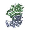
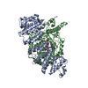
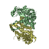
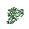
| Entry | Database: PDB / ID: 5uzr | ||||||
|---|---|---|---|---|---|---|---|
| Title | Crystal structure of citrate synthase from homo sapiens | ||||||
 Components Components | Citrate synthase, mitochondrial | ||||||
 Keywords Keywords | TRANSFERASE | ||||||
| Function / homology |  Function and homology informationcitrate (Si)-synthase / citrate (Si)-synthase activity / citrate metabolic process / Citric acid cycle (TCA cycle) / Mitochondrial protein import / Mitochondrial protein degradation / tricarboxylic acid cycle / carbohydrate metabolic process / mitochondrial matrix / mitochondrion ...citrate (Si)-synthase / citrate (Si)-synthase activity / citrate metabolic process / Citric acid cycle (TCA cycle) / Mitochondrial protein import / Mitochondrial protein degradation / tricarboxylic acid cycle / carbohydrate metabolic process / mitochondrial matrix / mitochondrion / RNA binding / extracellular exosome / nucleus Function and homology informationcitrate (Si)-synthase / citrate (Si)-synthase activity / citrate metabolic process / Citric acid cycle (TCA cycle) / Mitochondrial protein import / Mitochondrial protein degradation / tricarboxylic acid cycle / carbohydrate metabolic process / mitochondrial matrix / mitochondrion ...citrate (Si)-synthase / citrate (Si)-synthase activity / citrate metabolic process / Citric acid cycle (TCA cycle) / Mitochondrial protein import / Mitochondrial protein degradation / tricarboxylic acid cycle / carbohydrate metabolic process / mitochondrial matrix / mitochondrion / RNA binding / extracellular exosome / nucleusSimilarity search - Function | ||||||
| Biological species |  Homo sapiens (human) Homo sapiens (human) | ||||||
| Method | X-RAY DIFFRACTION / SYNCHROTRON / MOLECULAR REPLACEMENT / Resolution: 2.3 Å | ||||||
 Authors Authors | Schlachter, C. / Chruszcz, M. | ||||||
 Citation Citation | Journal: Biol.Chem. / Year: 2019 Title: Comparative studies of Aspergillus fumigatus 2-methylcitrate synthase and human citrate synthase. Authors: Schlachter, C.R. / Klapper, V. / Radford, T. / Chruszcz, M. | ||||||
| History |
|
- Structure visualization
| Structure viewer | Molecule: MolmilJmol/JSmol |
|---|
- Downloads & links
-Download
| PDBx/mmCIF format | 5uzr.cif.gz | 347.1 KB | Display | PDBx/mmCIF format |
|---|---|---|---|---|
| PDB format | pdb5uzr.ent.gz | 281.6 KB | Display | PDB format |
| PDBx/mmJSON format | 5uzr.json.gz | Tree view | PDBx/mmJSON format | |
| Others |  Other downloads Other downloads |
-Validation report
| Arichive directory | https://data.pdbj.org/pub/pdb/validation_reports/uz/5uzrftp://data.pdbj.org/pub/pdb/validation_reports/uz/5uzr | HTTPS FTP |
|---|
-Related structure data
| Related structure data | 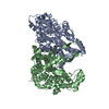 5uqoC 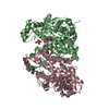 5uqqC 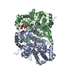 5uqrC 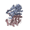 5uqsC 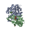 5uquC 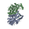 5uzpC 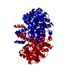 5uzqC  6bolC 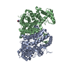 6bomC 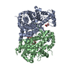 6bonC  6booC  6bopC C: citing same article ( |
|---|---|
| Similar structure data |


-Links
 PDBj
PDBj

- Assembly
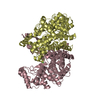
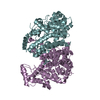
| Deposited unit | 
| ||||||||||||||||||
|---|---|---|---|---|---|---|---|---|---|---|---|---|---|---|---|---|---|---|---|
| 1 |
| ||||||||||||||||||
| Unit cell |
| ||||||||||||||||||
| Noncrystallographic symmetry (NCS) | NCS domain:
NCS domain segments: Component-ID: 0 / Ens-ID: 1 / Beg auth comp-ID: SER / Beg label comp-ID: SER / End auth comp-ID: ASP / End label comp-ID: ASP / Refine code: 0 / Auth seq-ID: 30 - 462 / Label seq-ID: 26 - 458
|
-Components
| #1: Protein | / Citrate (Si)-synthase Mass: 51736.848 Da / Num. of mol.: 2 / Fragment: residues 28-466 Source method: isolated from a genetically manipulated source Source: (gene. exp.) Homo sapiens (human) / Gene: CS / Production host:  Escherichia coli (E. coli) / References: UniProt: O75390, citrate (Si)-synthase Escherichia coli (E. coli) / References: UniProt: O75390, citrate (Si)-synthase#2: Chemical | Chloride  Mass: 35.453 Da / Num. of mol.: 2 / Source method: obtained synthetically / Formula: Cl Mass: 35.453 Da / Num. of mol.: 2 / Source method: obtained synthetically / Formula: Cl#3: Water | ChemComp-HOH / | Water Mass: 18.015 Da / Num. of mol.: 400 / Source method: isolated from a natural source / Formula: H2O Mass: 18.015 Da / Num. of mol.: 400 / Source method: isolated from a natural source / Formula: H2O |
|---|
-Experimental details
-Experiment
| Experiment | Method: X-RAY DIFFRACTION / Number of used crystals: 1 |
|---|
- Sample preparation
| Crystal | Density Matthews: 2.2 Å3/Da / Density % sol: 44.13 % |
|---|---|
| Crystal grow | Temperature: 298 K / Method: vapor diffusion, sitting drop / Details: 0.2 M Ammonium Acetate pH 7.1, 20% PEG3350 |
-Data collection
| Diffraction | Mean temperature: 100 K |
|---|---|
| Diffraction source | Source: SYNCHROTRON / Site: APS  / Beamline: 19-ID / Wavelength: 1 Å / Beamline: 19-ID / Wavelength: 1 Å |
| Detector | Type: ADSC QUANTUM 315r / Detector: CCD / Date: Jun 29, 2014 |
| Radiation | Protocol: SINGLE WAVELENGTH / Monochromatic (M) / Laue (L): M / Scattering type: x-ray |
| Radiation wavelength | Wavelength: 1 Å / Relative weight: 1 |
| Reflection | Resolution: 2.3→40 Å / Num. obs: 35831 / % possible obs: 96.15 % / Observed criterion σ(I): -3 / Redundancy: 2.1 % / Rmerge(I) obs: 0.078 / Rpim(I) all: 0.083 / Rrim(I) all: 0.123 / Rsym value: 0.078 / Net I/σ(I): 10.3 |
| Reflection shell | Resolution: 2.3→2.34 Å / Redundancy: 2.1 % / Rmerge(I) obs: 0.375 / Mean I/σ(I) obs: 2.18 / Num. unique obs: 1924 / CC1/2: 0.776 / Rpim(I) all: 0.372 / Rrim(I) all: 0.548 / Rsym value: 0.375 / % possible all: 97.7 |
- Processing
| Software |
| ||||||||||||||||||||||||||||||||||||||||||||||||||||||||||||||||||||||||||||||||||||||||||||||||||||||||||||||||||||||||||||||||||||||||||||||||||||||||||||||||||||||||||||||||||||||
|---|---|---|---|---|---|---|---|---|---|---|---|---|---|---|---|---|---|---|---|---|---|---|---|---|---|---|---|---|---|---|---|---|---|---|---|---|---|---|---|---|---|---|---|---|---|---|---|---|---|---|---|---|---|---|---|---|---|---|---|---|---|---|---|---|---|---|---|---|---|---|---|---|---|---|---|---|---|---|---|---|---|---|---|---|---|---|---|---|---|---|---|---|---|---|---|---|---|---|---|---|---|---|---|---|---|---|---|---|---|---|---|---|---|---|---|---|---|---|---|---|---|---|---|---|---|---|---|---|---|---|---|---|---|---|---|---|---|---|---|---|---|---|---|---|---|---|---|---|---|---|---|---|---|---|---|---|---|---|---|---|---|---|---|---|---|---|---|---|---|---|---|---|---|---|---|---|---|---|---|---|---|---|---|
| Refinement | Method to determine structure: MOLECULAR REPLACEMENT / Resolution: 2.3→40 Å / Cor.coef. Fo:Fc: 0.946 / Cor.coef. Fo:Fc free: 0.916 / SU B: 13.493 / SU ML: 0.16 / Cross valid method: THROUGHOUT / ESU R: 0.417 / ESU R Free: 0.229 / Details: HYDROGENS HAVE BEEN ADDED IN THE RIDING POSITIONS
| ||||||||||||||||||||||||||||||||||||||||||||||||||||||||||||||||||||||||||||||||||||||||||||||||||||||||||||||||||||||||||||||||||||||||||||||||||||||||||||||||||||||||||||||||||||||
| Solvent computation | Ion probe radii: 0.8 Å / Shrinkage radii: 0.8 Å / VDW probe radii: 1.2 Å | ||||||||||||||||||||||||||||||||||||||||||||||||||||||||||||||||||||||||||||||||||||||||||||||||||||||||||||||||||||||||||||||||||||||||||||||||||||||||||||||||||||||||||||||||||||||
| Displacement parameters | Biso mean: 26.323 Å2
| ||||||||||||||||||||||||||||||||||||||||||||||||||||||||||||||||||||||||||||||||||||||||||||||||||||||||||||||||||||||||||||||||||||||||||||||||||||||||||||||||||||||||||||||||||||||
| Refinement step | Cycle: 1 / Resolution: 2.3→40 Å
| ||||||||||||||||||||||||||||||||||||||||||||||||||||||||||||||||||||||||||||||||||||||||||||||||||||||||||||||||||||||||||||||||||||||||||||||||||||||||||||||||||||||||||||||||||||||
| Refine LS restraints |
|