 Movie
Movie Controller

[English] 日本語
 Yorodumi
- PDB-4cuc: Unravelling the multiple functions of the architecturally intrica... -
+ Open data

- Basic information
| Entry | Database: PDB / ID: 4cuc | |||||||||
|---|---|---|---|---|---|---|---|---|---|---|
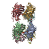
| Title | Unravelling the multiple functions of the architecturally intricate Streptococcus pneumoniae beta-galactosidase, BgaA. | |||||||||
 Components Components | BETA-GALACTOSIDASE | |||||||||
 Keywords Keywords | HYDROLASE | |||||||||
| Function / homology |  Function and homology informationbeta-galactosidase / carbohydrate catabolic process / beta-galactosidase activity / carbohydrate metabolic process / extracellular region Function and homology informationbeta-galactosidase / carbohydrate catabolic process / beta-galactosidase activity / carbohydrate metabolic process / extracellular regionSimilarity search - Function | |||||||||
| Biological species |   STREPTOCOCCUS PNEUMONIAE (bacteria) STREPTOCOCCUS PNEUMONIAE (bacteria) | |||||||||
| Method | X-RAY DIFFRACTION / SYNCHROTRON / MOLECULAR REPLACEMENT / Resolution: 2.2 Å | |||||||||
 Authors Authors | Singh, A.K. / Pluvinage, B. / Higgins, M.A. / Dalia, A.B. / Flynn, M. / Lloyd, A.R. / Weiser, J.N. / Stubbs, K.A. / Boraston, A.B. / King, S.J. | |||||||||
 Citation Citation | Journal: Plos Pathog. / Year: 2014 Title: Unravelling the Multiple Functions of the Architecturally Intricate Streptococcus Pneumoniae Beta-Galactosidase, BgaA. Authors: Singh, A.K. / Pluvinage, B. / Higgins, M.A. / Dalia, A.B. / Woodiga, S.A. / Flynn, M. / Lloyd, A.R. / Weiser, J.N. / Stubbs, K.A. / Boraston, A.B. / King, S.J. | |||||||||
| History |
| |||||||||
| Remark 700 | SHEET DETERMINATION METHOD: DSSP THE SHEETS PRESENTED AS "AJ" IN EACH CHAIN ON SHEET RECORDS BELOW ... SHEET DETERMINATION METHOD: DSSP THE SHEETS PRESENTED AS "AJ" IN EACH CHAIN ON SHEET RECORDS BELOW IS ACTUALLY AN 8-STRANDED BARREL THIS IS REPRESENTED BY A 9-STRANDED SHEET IN WHICH THE FIRST AND LAST STRANDS ARE IDENTICAL. |
- Structure visualization
| Structure viewer | Molecule: MolmilJmol/JSmol |
|---|
- Downloads & links
-Download
| PDBx/mmCIF format | 4cuc.cif.gz | 208.6 KB | Display | PDBx/mmCIF format |
|---|---|---|---|---|
| PDB format | pdb4cuc.ent.gz | 163.1 KB | Display | PDB format |
| PDBx/mmJSON format | 4cuc.json.gz | Tree view | PDBx/mmJSON format | |
| Others |  Other downloads Other downloads |
-Validation report
| Arichive directory | https://data.pdbj.org/pub/pdb/validation_reports/cu/4cucftp://data.pdbj.org/pub/pdb/validation_reports/cu/4cuc | HTTPS FTP |
|---|
-Related structure data
| Related structure data | 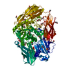 4cu6C 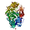 4cu7C 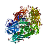 4cu8C 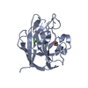 4cu9C  4cuaC  4cubC C: citing same article ( |
|---|---|
| Similar structure data |


-Links
 PDBj
PDBj

- Assembly
| Deposited unit | 
| ||||||||
|---|---|---|---|---|---|---|---|---|---|
| 1 | 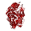
| ||||||||
| Unit cell |
| ||||||||
| Components on special symmetry positions |
|
-Components
| #1: Protein | / GLYCOSIDE HYDROLASE 2 Mass: 96324.133 Da / Num. of mol.: 1 / Fragment: CATALYTIC MODULE, RESIDUES 137-985 / Mutation: YES Source method: isolated from a genetically manipulated source Source: (gene. exp.) STREPTOCOCCUS PNEUMONIAE (bacteria) / Strain: TIGR4 / Production host: ESCHERICHIA COLI (E. coli) / References: UniProt: I6L8R4, UniProt: A0A0H2UP19*PLUS | ||||
|---|---|---|---|---|---|
| #2: Polysaccharide | beta-D-galactopyranose-(1-4)-2-acetamido-2-deoxy-beta-D-glucopyranose / Mass: 383.349 Da / Num. of mol.: 1 Source method: isolated from a genetically manipulated source | ||||
| #3: Chemical | ChemComp-SO4 / Sulfate  Mass: 96.063 Da / Num. of mol.: 6 / Source method: obtained synthetically / Formula: SO4 Mass: 96.063 Da / Num. of mol.: 6 / Source method: obtained synthetically / Formula: SO4#4: Chemical | ChemComp-MPD / ( 2-Methyl-2,4-pentanediol  Mass: 118.174 Da / Num. of mol.: 5 / Source method: obtained synthetically / Formula: C6H14O2 / Comment: precipitant*YM Mass: 118.174 Da / Num. of mol.: 5 / Source method: obtained synthetically / Formula: C6H14O2 / Comment: precipitant*YM#5: Water | ChemComp-HOH / | Water Mass: 18.015 Da / Num. of mol.: 800 / Source method: isolated from a natural source / Formula: H2O Mass: 18.015 Da / Num. of mol.: 800 / Source method: isolated from a natural source / Formula: H2O |
-Experimental details
-Experiment
| Experiment | Method: X-RAY DIFFRACTION / Number of used crystals: 1 |
|---|
- Sample preparation
| Crystal | Density Matthews: 3.92 Å3/Da / Density % sol: 68.65 % / Description: NONE |
|---|
-Data collection
| Diffraction | Mean temperature: 100 K |
|---|---|
| Diffraction source | Source: SYNCHROTRON / Site: CLSI  / Beamline: 08ID-1 / Type: CLSI / Wavelength: 0.97949 / Beamline: 08ID-1 / Type: CLSI / Wavelength: 0.97949 |
| Detector | Type: MARRESEARCH / Detector: CCD |
| Radiation | Protocol: SINGLE WAVELENGTH / Monochromatic (M) / Laue (L): M / Scattering type: x-ray |
| Radiation wavelength | Wavelength: 0.97949 Å / Relative weight: 1 |
| Reflection | Resolution: 2.2→52.49 Å / Num. obs: 78624 / % possible obs: 100 % / Observed criterion σ(I): 2 / Redundancy: 9.8 % / Rmerge(I) obs: 0.2 / Net I/σ(I): 10.1 |
| Reflection shell | Resolution: 2.2→2.32 Å / Redundancy: 9.8 % / Rmerge(I) obs: 0.64 / Mean I/σ(I) obs: 5.6 / % possible all: 100 |
- Processing
| Software |
| ||||||||||||||||||||||||||||||||||||||||||||||||||||||||||||||||||||||||||||||||||||||||||||||||||||||||||||||||||||||||||||||||||||||||||||||||||||||||||||||||||||||||||||||||||||||
|---|---|---|---|---|---|---|---|---|---|---|---|---|---|---|---|---|---|---|---|---|---|---|---|---|---|---|---|---|---|---|---|---|---|---|---|---|---|---|---|---|---|---|---|---|---|---|---|---|---|---|---|---|---|---|---|---|---|---|---|---|---|---|---|---|---|---|---|---|---|---|---|---|---|---|---|---|---|---|---|---|---|---|---|---|---|---|---|---|---|---|---|---|---|---|---|---|---|---|---|---|---|---|---|---|---|---|---|---|---|---|---|---|---|---|---|---|---|---|---|---|---|---|---|---|---|---|---|---|---|---|---|---|---|---|---|---|---|---|---|---|---|---|---|---|---|---|---|---|---|---|---|---|---|---|---|---|---|---|---|---|---|---|---|---|---|---|---|---|---|---|---|---|---|---|---|---|---|---|---|---|---|---|---|
| Refinement | Method to determine structure: MOLECULAR REPLACEMENT / Resolution: 2.2→117.34 Å / Cor.coef. Fo:Fc: 0.96 / Cor.coef. Fo:Fc free: 0.935 / SU B: 3.45 / SU ML: 0.089 / Cross valid method: THROUGHOUT / ESU R: 0.148 / ESU R Free: 0.14 / Stereochemistry target values: MAXIMUM LIKELIHOOD Details: HYDROGENS HAVE BEEN ADDED IN THE RIDING POSITIONS. HYDROGENS HAVE BEEN USED IF PRESENT IN THE INPUT U VALUES REFINED INDIVIDUALLY
| ||||||||||||||||||||||||||||||||||||||||||||||||||||||||||||||||||||||||||||||||||||||||||||||||||||||||||||||||||||||||||||||||||||||||||||||||||||||||||||||||||||||||||||||||||||||
| Solvent computation | Ion probe radii: 0.8 Å / Shrinkage radii: 0.8 Å / VDW probe radii: 1.2 Å / Solvent model: MASK | ||||||||||||||||||||||||||||||||||||||||||||||||||||||||||||||||||||||||||||||||||||||||||||||||||||||||||||||||||||||||||||||||||||||||||||||||||||||||||||||||||||||||||||||||||||||
| Displacement parameters | Biso mean: 23.354 Å2
| ||||||||||||||||||||||||||||||||||||||||||||||||||||||||||||||||||||||||||||||||||||||||||||||||||||||||||||||||||||||||||||||||||||||||||||||||||||||||||||||||||||||||||||||||||||||
| Refinement step | Cycle: LAST / Resolution: 2.2→117.34 Å
| ||||||||||||||||||||||||||||||||||||||||||||||||||||||||||||||||||||||||||||||||||||||||||||||||||||||||||||||||||||||||||||||||||||||||||||||||||||||||||||||||||||||||||||||||||||||
| Refine LS restraints |
|