 Movie
Movie Controller


+ Open data

- Basic information
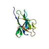
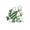
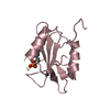
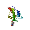
| Entry | Database: PDB / ID: 5uzm | ||||||
|---|---|---|---|---|---|---|---|
| Title | Crystal structure of Glorund qRRM2 domain | ||||||
 Components Components | AT27789p | ||||||
 Keywords Keywords |  RNA BINDING PROTEIN / quasi-RNA recognition motif / qRRM RNA BINDING PROTEIN / quasi-RNA recognition motif / qRRM | ||||||
| Function / homology |  Function and homology information Function and homology informationmaternal specification of dorsal/ventral axis, oocyte, germ-line encoded / intracellular mRNA localization involved in pattern specification process / Processing of Capped Intron-Containing Pre-mRNA / mRNA Splicing - Major Pathway / pole plasm oskar mRNA localization / regulation of RNA splicing / chromosome organization / wound healing / regulation of translation / ribonucleoprotein complex ...maternal specification of dorsal/ventral axis, oocyte, germ-line encoded / intracellular mRNA localization involved in pattern specification process / Processing of Capped Intron-Containing Pre-mRNA / mRNA Splicing - Major Pathway / pole plasm oskar mRNA localization / regulation of RNA splicing / chromosome organization / wound healing / regulation of translation / ribonucleoprotein complex / mRNA binding / protein-containing complex / RNA binding / nucleoplasmSimilarity search - Function | ||||||
| Biological species |  Drosophila melanogaster (fruit fly) Drosophila melanogaster (fruit fly) | ||||||
| Method | X-RAY DIFFRACTION / SYNCHROTRON / MOLECULAR REPLACEMENT / Resolution: 1.552 Å | ||||||
 Authors Authors | Teramoto, T. / Hall, T.M.T. | ||||||
 Citation Citation | Journal: Cell Rep / Year: 2017 Title: The Drosophila hnRNP F/H Homolog Glorund Uses Two Distinct RNA-Binding Modes to Diversify Target Recognition. Authors: Tamayo, J.V. / Teramoto, T. / Chatterjee, S. / Hall, T.M. / Gavis, E.R. | ||||||
| History |
|
- Structure visualization
| Structure viewer | Molecule: MolmilJmol/JSmol |
|---|
- Downloads & links
-Download
| PDBx/mmCIF format | 5uzm.cif.gz | 53.1 KB | Display | PDBx/mmCIF format |
|---|---|---|---|---|
| PDB format | pdb5uzm.ent.gz | 37.1 KB | Display | PDB format |
| PDBx/mmJSON format | 5uzm.json.gz | Tree view | PDBx/mmJSON format | |
| Others |  Other downloads Other downloads |
-Validation report
| Arichive directory | https://data.pdbj.org/pub/pdb/validation_reports/uz/5uzmftp://data.pdbj.org/pub/pdb/validation_reports/uz/5uzm | HTTPS FTP |
|---|
-Related structure data
| Related structure data | 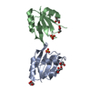 5uzgC 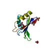 5uznC 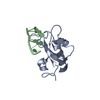 2kg0S C: citing same article ( S: Starting model for refinement |
|---|---|
| Similar structure data |


-Links
 PDBj
PDBj- Assembly
| Deposited unit | 
| ||||||||
|---|---|---|---|---|---|---|---|---|---|
| 1 | 
| ||||||||
| 2 | 
| ||||||||
| Unit cell |
|
-Components
| #1: Protein | Mass: 11024.400 Da / Num. of mol.: 2 / Fragment: residues 142-234 Source method: isolated from a genetically manipulated source Source: (gene. exp.) Drosophila melanogaster (fruit fly) / Gene: glo, CG6946, Dmel_CG6946 / Production host:  Escherichia coli (E. coli) / References: UniProt: Q9VGH5 Escherichia coli (E. coli) / References: UniProt: Q9VGH5#2: Water | ChemComp-HOH / | Water Mass: 18.015 Da / Num. of mol.: 127 / Source method: isolated from a natural source / Formula: H2O Mass: 18.015 Da / Num. of mol.: 127 / Source method: isolated from a natural source / Formula: H2O |
|---|
-Experimental details
-Experiment
| Experiment | Method: X-RAY DIFFRACTION / Number of used crystals: 1 |
|---|
- Sample preparation
| Crystal | Density Matthews: 2.08 Å3/Da / Density % sol: 40.85 % |
|---|---|
| Crystal grow | Temperature: 293 K / Method: vapor diffusion, hanging drop / Details: 0.2 M ammonium acetate, 20% v/v PEG 3350 |
-Data collection
| Diffraction | Mean temperature: 100 K |
|---|---|
| Diffraction source | Source: SYNCHROTRON / Site: APS  / Beamline: 22-ID / Wavelength: 1 Å / Beamline: 22-ID / Wavelength: 1 Å |
| Detector | Type: MARMOSAIC 300 mm CCD / Detector: CCD / Date: Feb 24, 2012 |
| Radiation | Protocol: SINGLE WAVELENGTH / Monochromatic (M) / Laue (L): M / Scattering type: x-ray |
| Radiation wavelength | Wavelength: 1 Å / Relative weight: 1 |
| Reflection | Resolution: 1.55→50 Å / Num. obs: 23996 / % possible obs: 93.4 % / Redundancy: 3.4 % / Rmerge(I) obs: 0.05 / Net I/σ(I): 34.6 |
- Processing
| Software |
| ||||||||||||||||||||||||||||||||||||||||||||||||||||||||||||||||||||||
|---|---|---|---|---|---|---|---|---|---|---|---|---|---|---|---|---|---|---|---|---|---|---|---|---|---|---|---|---|---|---|---|---|---|---|---|---|---|---|---|---|---|---|---|---|---|---|---|---|---|---|---|---|---|---|---|---|---|---|---|---|---|---|---|---|---|---|---|---|---|---|---|
| Refinement | Method to determine structure: MOLECULAR REPLACEMENT Starting model: 2KG0 Resolution: 1.552→32.542 Å / SU ML: 0.2 / Cross valid method: FREE R-VALUE / σ(F): 1.24 / Phase error: 25.94
| ||||||||||||||||||||||||||||||||||||||||||||||||||||||||||||||||||||||
| Solvent computation | Shrinkage radii: 0.9 Å / VDW probe radii: 1.11 Å | ||||||||||||||||||||||||||||||||||||||||||||||||||||||||||||||||||||||
| Refinement step | Cycle: LAST / Resolution: 1.552→32.542 Å
| ||||||||||||||||||||||||||||||||||||||||||||||||||||||||||||||||||||||
| Refine LS restraints |
| ||||||||||||||||||||||||||||||||||||||||||||||||||||||||||||||||||||||
| LS refinement shell |
|
