 Movie
Movie Controller

[English] 日本語
 Yorodumi
- PDB-5t6o: Structure of the catalytic domain of the class I polyhydroxybutyr... -
+ Open data

- Basic information
| Entry | Database: PDB / ID: 5t6o | |||||||||
|---|---|---|---|---|---|---|---|---|---|---|
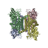
| Title | Structure of the catalytic domain of the class I polyhydroxybutyrate synthase from Cupriavidus necator | |||||||||
 Components Components | Poly-beta-hydroxybuterate polymerase | |||||||||
 Keywords Keywords |  BIOSYNTHETIC PROTEIN / polyhydroxybutyrate synthase / PhaC / alpha/beta hydrolase fold / nontemplate-dependent polymerization / TRANSFERASE BIOSYNTHETIC PROTEIN / polyhydroxybutyrate synthase / PhaC / alpha/beta hydrolase fold / nontemplate-dependent polymerization / TRANSFERASE | |||||||||
| Function / homology |  Function and homology information Function and homology informationpoly-hydroxybutyrate biosynthetic process / acyltransferase activity / Transferases; Acyltransferases; Transferring groups other than aminoacyl groups / cytoplasmSimilarity search - Function | |||||||||
| Biological species |  Cupriavidus necator (bacteria) Cupriavidus necator (bacteria) | |||||||||
| Method | X-RAY DIFFRACTION / SYNCHROTRON / MAD / Resolution: 1.8 Å | |||||||||
 Authors Authors | Wittenborn, E.C. / Jost, M. / Drennan, C.L. | |||||||||
| Funding support |  United States, 2items United States, 2items
| |||||||||
 Citation Citation | Journal: J.Biol.Chem. / Year: 2016 Title: Structure of the Catalytic Domain of the Class I Polyhydroxybutyrate Synthase from Cupriavidus necator. Authors: Wittenborn, E.C. / Jost, M. / Wei, Y. / Stubbe, J. / Drennan, C.L. | |||||||||
| History |
|
- Structure visualization
| Structure viewer | Molecule: MolmilJmol/JSmol |
|---|
- Downloads & links
-Download
| PDBx/mmCIF format | 5t6o.cif.gz | 162.1 KB | Display | PDBx/mmCIF format |
|---|---|---|---|---|
| PDB format | pdb5t6o.ent.gz | 132.2 KB | Display | PDB format |
| PDBx/mmJSON format | 5t6o.json.gz | Tree view | PDBx/mmJSON format | |
| Others |  Other downloads Other downloads |
-Validation report
| Arichive directory | https://data.pdbj.org/pub/pdb/validation_reports/t6/5t6oftp://data.pdbj.org/pub/pdb/validation_reports/t6/5t6o | HTTPS FTP |
|---|
-Related structure data
| Similar structure data |
|---|

-Links
 PDBj
PDBj


- Assembly
| Deposited unit | 
| ||||||||
|---|---|---|---|---|---|---|---|---|---|
| 1 | 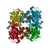
| ||||||||
| Unit cell |
| ||||||||
| Components on special symmetry positions |
|
-Components
| #1: Protein | Mass: 42635.242 Da / Num. of mol.: 1 / Fragment: UNP residues 201-589 Source method: isolated from a genetically manipulated source Source: (gene. exp.) Cupriavidus necator (bacteria) / Gene: phaC / Production host: Escherichia coli (E. coli) / References: UniProt: D3UAK8, UniProt: P23608*PLUS | ||
|---|---|---|---|
| #2: Chemical | ChemComp-SO4 / Sulfate  Mass: 96.063 Da / Num. of mol.: 4 / Source method: obtained synthetically / Formula: SO4 Mass: 96.063 Da / Num. of mol.: 4 / Source method: obtained synthetically / Formula: SO4#3: Water | ChemComp-HOH / | Water Mass: 18.015 Da / Num. of mol.: 283 / Source method: isolated from a natural source / Formula: H2O Mass: 18.015 Da / Num. of mol.: 283 / Source method: isolated from a natural source / Formula: H2O |
-Experimental details
-Experiment
| Experiment | Method: X-RAY DIFFRACTION / Number of used crystals: 1 |
|---|
- Sample preparation
| Crystal | Density Matthews: 2.6 Å3/Da / Density % sol: 52.63 % |
|---|---|
| Crystal grow | Temperature: 298 K / Method: vapor diffusion, hanging drop / pH: 7 / Details: 0.9 M ammonium sulfate, 0.1 M HEPES pH 7 |
-Data collection
| Diffraction | Mean temperature: 100 K |
|---|---|
| Diffraction source | Source: SYNCHROTRON / Site: APS / Beamline: 24-ID-C / Wavelength: 0.9792 Å |
| Detector | Type: DECTRIS PILATUS 6M-F / Detector: PIXEL / Date: Aug 23, 2015 |
| Radiation | Protocol: SINGLE WAVELENGTH / Monochromatic (M) / Laue (L): M / Scattering type: x-ray |
| Radiation wavelength | Wavelength: 0.9792 Å / Relative weight: 1 |
| Reflection | Resolution: 1.8→100 Å / Num. obs: 40649 / % possible obs: 99.9 % / Redundancy: 6.6 % / CC1/2: 1 / Rsym value: 0.046 / Net I/σ(I): 25.76 |
| Reflection shell | Resolution: 1.8→1.84 Å / Redundancy: 6.7 % / Mean I/σ(I) obs: 2.24 / CC1/2: 0.762 / % possible all: 100 |
- Processing
| Software |
| ||||||||||||||||||||||||||||||||||||||||||||||||||||||||||||||||||||||||||||||||||||||||||||||||||||||||||||||||
|---|---|---|---|---|---|---|---|---|---|---|---|---|---|---|---|---|---|---|---|---|---|---|---|---|---|---|---|---|---|---|---|---|---|---|---|---|---|---|---|---|---|---|---|---|---|---|---|---|---|---|---|---|---|---|---|---|---|---|---|---|---|---|---|---|---|---|---|---|---|---|---|---|---|---|---|---|---|---|---|---|---|---|---|---|---|---|---|---|---|---|---|---|---|---|---|---|---|---|---|---|---|---|---|---|---|---|---|---|---|---|---|---|---|
| Refinement | Method to determine structure: MAD / Resolution: 1.8→73.95 Å / SU ML: 0.2 / Cross valid method: FREE R-VALUE / σ(F): 1.36 / Phase error: 20.56
| ||||||||||||||||||||||||||||||||||||||||||||||||||||||||||||||||||||||||||||||||||||||||||||||||||||||||||||||||
| Solvent computation | Shrinkage radii: 0.9 Å / VDW probe radii: 1.11 Å | ||||||||||||||||||||||||||||||||||||||||||||||||||||||||||||||||||||||||||||||||||||||||||||||||||||||||||||||||
| Refinement step | Cycle: LAST / Resolution: 1.8→73.95 Å
| ||||||||||||||||||||||||||||||||||||||||||||||||||||||||||||||||||||||||||||||||||||||||||||||||||||||||||||||||
| Refine LS restraints |
| ||||||||||||||||||||||||||||||||||||||||||||||||||||||||||||||||||||||||||||||||||||||||||||||||||||||||||||||||
| LS refinement shell |
| ||||||||||||||||||||||||||||||||||||||||||||||||||||||||||||||||||||||||||||||||||||||||||||||||||||||||||||||||
| Refinement TLS params. | Method: refined / Origin x: 86.6891 Å / Origin y: 28.4958 Å / Origin z: 18.1841 Å
| ||||||||||||||||||||||||||||||||||||||||||||||||||||||||||||||||||||||||||||||||||||||||||||||||||||||||||||||||
| Refinement TLS group | Selection details: (chain 'A' and resid 201 through 589) |
