 Movie
Movie Controller


+ Open data

- Basic information
| Entry | Database: PDB / ID: 5t12 | ||||||
|---|---|---|---|---|---|---|---|
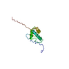
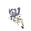
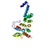
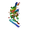
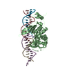
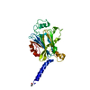
| Title | N-terminal domain of Enzyme 1 - Nitrogen | ||||||
 Components Components | Phosphoenolpyruvate--protein phosphotransferase | ||||||
 Keywords Keywords |  TRANSFERASE / PTSNtr / phosphotransfer TRANSFERASE / PTSNtr / phosphotransfer | ||||||
| Function / homology |  Function and homology informationphosphoenolpyruvate-protein phosphotransferase / phosphoenolpyruvate-protein phosphotransferase activity / phosphoenolpyruvate-dependent sugar phosphotransferase system / response to organonitrogen compound / kinase activity / phosphorylation / metal ion binding / cytoplasm Function and homology informationphosphoenolpyruvate-protein phosphotransferase / phosphoenolpyruvate-protein phosphotransferase activity / phosphoenolpyruvate-dependent sugar phosphotransferase system / response to organonitrogen compound / kinase activity / phosphorylation / metal ion binding / cytoplasmSimilarity search - Function | ||||||
| Biological species |  Escherichia coli (E. coli) Escherichia coli (E. coli) | ||||||
| Method | X-RAY DIFFRACTION / SYNCHROTRON / MOLECULAR REPLACEMENT / Resolution: 2.299 Å | ||||||
 Authors Authors | Stanley, A.M. / Botos, I. / Buchanan, S.K. | ||||||
 Citation Citation | Journal: Structure / Year: 2016 Title: Structure of the NPr:EIN(Ntr) Complex: Mechanism for Specificity in Paralogous Phosphotransferase Systems. Authors: Strickland, M. / Stanley, A.M. / Wang, G. / Botos, I. / Schwieters, C.D. / Buchanan, S.K. / Peterkofsky, A. / Tjandra, N. | ||||||
| History |
|
- Structure visualization
| Structure viewer | Molecule: MolmilJmol/JSmol |
|---|
- Downloads & links
-Download
| PDBx/mmCIF format | 5t12.cif.gz | 61.8 KB | Display | PDBx/mmCIF format |
|---|---|---|---|---|
| PDB format | pdb5t12.ent.gz | 43.6 KB | Display | PDB format |
| PDBx/mmJSON format | 5t12.json.gz | Tree view | PDBx/mmJSON format | |
| Others |  Other downloads Other downloads |
-Validation report
| Arichive directory | https://data.pdbj.org/pub/pdb/validation_reports/t1/5t12ftp://data.pdbj.org/pub/pdb/validation_reports/t1/5t12 | HTTPS FTP |
|---|
-Related structure data
| Related structure data | 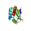 5t1nC  5t1oC  1zymS S: Starting model for refinement C: citing same article ( |
|---|---|
| Similar structure data |

-Links
 PDBj
PDBj


- Assembly
| Deposited unit | 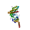
| ||||||||
|---|---|---|---|---|---|---|---|---|---|
| 1 |
| ||||||||
| Unit cell |
|
-Components
| #1: Protein | Mass: 28323.018 Da / Num. of mol.: 1 / Fragment: residues 170-424 / Mutation: H356Q Source method: isolated from a genetically manipulated source Source: (gene. exp.) Escherichia coli (E. coli)Gene: ptsP, AC789_1c31510, ACU90_23270, AML37_15090, AWH59_07225, WQ89_09920 Production host: Escherichia coli (E. coli)References: UniProt: A0A0E1LBH7, UniProt: P37177*PLUS, phosphoenolpyruvate-protein phosphotransferase | ||
|---|---|---|---|
| #2: Chemical | ChemComp-IOD / Iodide  Mass: 126.904 Da / Num. of mol.: 5 / Source method: obtained synthetically / Formula: I Mass: 126.904 Da / Num. of mol.: 5 / Source method: obtained synthetically / Formula: I#3: Water | ChemComp-HOH / | Water Mass: 18.015 Da / Num. of mol.: 41 / Source method: isolated from a natural source / Formula: H2O Mass: 18.015 Da / Num. of mol.: 41 / Source method: isolated from a natural source / Formula: H2O |
-Experimental details
-Experiment
| Experiment | Method: X-RAY DIFFRACTION / Number of used crystals: 1 |
|---|
- Sample preparation
| Crystal | Density Matthews: 2.64 Å3/Da / Density % sol: 53.49 % |
|---|---|
| Crystal grow | Temperature: 298.15 K / Method: vapor diffusion, hanging drop Details: 1.0M Potassium/Sodium tartrate 0.1M imidazole 8.0 0.2M NaCl 1.0M sodium iodide |
-Data collection
| Diffraction | Mean temperature: 90 K |
|---|---|
| Diffraction source | Source: SYNCHROTRON / Site: APS  / Beamline: 22-ID / Wavelength: 1 Å / Beamline: 22-ID / Wavelength: 1 Å |
| Detector | Type: MARMOSAIC 300 mm CCD / Detector: CCD / Date: Mar 21, 2012 |
| Radiation | Protocol: SINGLE WAVELENGTH / Monochromatic (M) / Laue (L): M / Scattering type: x-ray |
| Radiation wavelength | Wavelength: 1 Å / Relative weight: 1 |
| Reflection | Resolution: 2.299→33.221 Å / Num. obs: 11880 / % possible obs: 96.2 % / Redundancy: 4.3 % / Rmerge(I) obs: 0.112 / Net I/σ(I): 12.8 |
| Reflection shell | Resolution: 2.299→2.38 Å / Redundancy: 2.3 % / Rmerge(I) obs: 0.509 / Mean I/σ(I) obs: 1.48 / % possible all: 90.3 |
- Processing
| Software |
| |||||||||||||||||||||||||||||||||||
|---|---|---|---|---|---|---|---|---|---|---|---|---|---|---|---|---|---|---|---|---|---|---|---|---|---|---|---|---|---|---|---|---|---|---|---|---|
| Refinement | Method to determine structure: MOLECULAR REPLACEMENT Starting model: 1ZYM Resolution: 2.299→33.221 Å / SU ML: 0.36 / Cross valid method: FREE R-VALUE / σ(F): 0 / Phase error: 30.31
| |||||||||||||||||||||||||||||||||||
| Solvent computation | Shrinkage radii: 0.9 Å / VDW probe radii: 1.11 Å | |||||||||||||||||||||||||||||||||||
| Refinement step | Cycle: LAST / Resolution: 2.299→33.221 Å
| |||||||||||||||||||||||||||||||||||
| Refine LS restraints |
| |||||||||||||||||||||||||||||||||||
| LS refinement shell |
|
