 Movie
Movie Controller


+ Open data

- Basic information
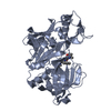
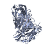
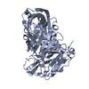
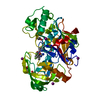
| Entry | Database: PDB / ID: 5qd4 | ||||||
|---|---|---|---|---|---|---|---|
| Title | Crystal structure of BACE complex with BMC023 | ||||||
 Components Components | Beta-secretase 1 | ||||||
 Keywords Keywords | HYDROLASE / Hydroloase / D3R / BACE / Ligand Docking | ||||||
| Function / homology |  Function and homology informationmemapsin 2 / Golgi-associated vesicle lumen / signaling receptor ligand precursor processing / beta-aspartyl-peptidase activity / amyloid precursor protein catabolic process / amyloid-beta formation / membrane protein ectodomain proteolysis / cellular response to manganese ion / prepulse inhibition / detection of mechanical stimulus involved in sensory perception of pain ...memapsin 2 / Golgi-associated vesicle lumen / signaling receptor ligand precursor processing / beta-aspartyl-peptidase activity / amyloid precursor protein catabolic process / amyloid-beta formation / membrane protein ectodomain proteolysis / cellular response to manganese ion / prepulse inhibition / detection of mechanical stimulus involved in sensory perception of pain / amyloid-beta metabolic process / cellular response to copper ion / hippocampal mossy fiber to CA3 synapse / presynaptic modulation of chemical synaptic transmission / multivesicular body / response to lead ion / trans-Golgi network / protein processing / recycling endosome / cellular response to amyloid-beta / positive regulation of neuron apoptotic process / synaptic vesicle / late endosome / amyloid-beta binding / peptidase activity / endopeptidase activity / amyloid fibril formation / lysosome / aspartic-type endopeptidase activity / early endosome / endosome membrane / endosome / membrane raft / Amyloid fiber formation / axon / endoplasmic reticulum lumen / neuronal cell body / dendrite / Golgi apparatus / enzyme binding / cell surface / proteolysis / membrane / plasma membrane Function and homology informationmemapsin 2 / Golgi-associated vesicle lumen / signaling receptor ligand precursor processing / beta-aspartyl-peptidase activity / amyloid precursor protein catabolic process / amyloid-beta formation / membrane protein ectodomain proteolysis / cellular response to manganese ion / prepulse inhibition / detection of mechanical stimulus involved in sensory perception of pain ...memapsin 2 / Golgi-associated vesicle lumen / signaling receptor ligand precursor processing / beta-aspartyl-peptidase activity / amyloid precursor protein catabolic process / amyloid-beta formation / membrane protein ectodomain proteolysis / cellular response to manganese ion / prepulse inhibition / detection of mechanical stimulus involved in sensory perception of pain / amyloid-beta metabolic process / cellular response to copper ion / hippocampal mossy fiber to CA3 synapse / presynaptic modulation of chemical synaptic transmission / multivesicular body / response to lead ion / trans-Golgi network / protein processing / recycling endosome / cellular response to amyloid-beta / positive regulation of neuron apoptotic process / synaptic vesicle / late endosome / amyloid-beta binding / peptidase activity / endopeptidase activity / amyloid fibril formation / lysosome / aspartic-type endopeptidase activity / early endosome / endosome membrane / endosome / membrane raft / Amyloid fiber formation / axon / endoplasmic reticulum lumen / neuronal cell body / dendrite / Golgi apparatus / enzyme binding / cell surface / proteolysis / membrane / plasma membraneSimilarity search - Function | ||||||
| Biological species |  Homo sapiens (human) Homo sapiens (human) | ||||||
| Method | X-RAY DIFFRACTION / SYNCHROTRON / MOLECULAR REPLACEMENT / molecular replacement / Resolution: 2.112 Å | ||||||
| Model details | Structures for D3R docking challenge | ||||||
 Authors Authors | Rondeau, J.M. / Shao, C. / Yang, H. / Burley, S.K. | ||||||
 Citation Citation | Journal: J.Comput.Aided Mol.Des. / Year: 2020 Title: D3R grand challenge 4: blind prediction of protein-ligand poses, affinity rankings, and relative binding free energies. Authors: Parks, C.D. / Gaieb, Z. / Chiu, M. / Yang, H. / Shao, C. / Walters, W.P. / Jansen, J.M. / McGaughey, G. / Lewis, R.A. / Bembenek, S.D. / Ameriks, M.K. / Mirzadegan, T. / Burley, S.K. / Amaro, R.E. / Gilson, M.K. #1: Journal: J.Med.Chem. / Year: 2006Title: Structure-based design and synthesis of macroheterocyclic peptidomimetic inhibitors of the aspartic protease beta-site amyloid precursor protein cleaving enzyme (BACE). Authors: Hanessian, S. / Yang, G. / Rondeau, J.M. / Neumann, U. / Betschart, C. / Tintelnot-Blomley, M. | ||||||
| History |
|
- Structure visualization
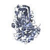
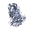
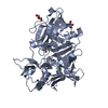
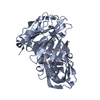
| Structure viewer | Molecule: MolmilJmol/JSmol |
|---|
- Downloads & links
-Download
| PDBx/mmCIF format | 5qd4.cif.gz | 460.4 KB | Display | PDBx/mmCIF format |
|---|---|---|---|---|
| PDB format | pdb5qd4.ent.gz | 393.7 KB | Display | PDB format |
| PDBx/mmJSON format | 5qd4.json.gz | Tree view | PDBx/mmJSON format | |
| Others |  Other downloads Other downloads |
-Validation report
| Arichive directory | https://data.pdbj.org/pub/pdb/validation_reports/qd/5qd4ftp://data.pdbj.org/pub/pdb/validation_reports/qd/5qd4 | HTTPS FTP |
|---|
-Group deposition
| ID | G_1002044 (26 entries) |
|---|---|
| Title | Crystal Structures of Beta-Secretase 1 with Bound Ligands |
| Type | undefined |
| Description | Crystal Structures of Complexes of Beta-Secretase 1 and Ligands |


-Related structure data
| Similar structure data |
|---|


-Links
 PDBj
PDBj

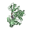
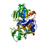
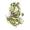
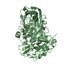
- Assembly
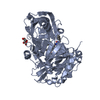
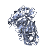
| Deposited unit | 
| ||||||||
|---|---|---|---|---|---|---|---|---|---|
| 1 | 
| ||||||||
| 2 | 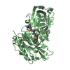
| ||||||||
| 3 | 
| ||||||||
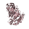
| Unit cell |
|
-Components
| #1: Protein | / Aspartyl protease 2 / Asp 2 / Beta-site amyloid precursor protein cleaving enzyme 1 / Beta-site APP ...Aspartyl protease 2 / Asp 2 / Beta-site amyloid precursor protein cleaving enzyme 1 / Beta-site APP cleaving enzyme 1 / Memapsin-2 / Membrane-associated aspartic protease 2 Mass: 44777.336 Da / Num. of mol.: 3 Source method: isolated from a genetically manipulated source Source: (gene. exp.) Homo sapiens (human) / Gene: BACE1, BACE, KIAA1149 / Production host:  Escherichia coli (E. coli) / References: UniProt: P56817, memapsin 2 Escherichia coli (E. coli) / References: UniProt: P56817, memapsin 2#2: Chemical |   Mass: 673.928 Da / Num. of mol.: 3 / Source method: obtained synthetically / Formula: C31H55N5O7S2 Mass: 673.928 Da / Num. of mol.: 3 / Source method: obtained synthetically / Formula: C31H55N5O7S2#3: Water | ChemComp-HOH / | Water Mass: 18.015 Da / Num. of mol.: 219 / Source method: isolated from a natural source / Formula: H2O Mass: 18.015 Da / Num. of mol.: 219 / Source method: isolated from a natural source / Formula: H2O |
|---|
-Experimental details
-Experiment
| Experiment | Method: X-RAY DIFFRACTION / Number of used crystals: 1 |
|---|
- Sample preparation
| Crystal | Density Matthews: 3.12 Å3/Da / Density % sol: 60.64 % |
|---|---|
| Crystal grow | Temperature: 292 K / Method: hanging drop Details: CRYSTALS WERE GROWN AT 19C BY VAPOUR DIFFUSION IN HANGING DROPS FROM 1.0M AMMONIUM PHOSPHATE, 0.1M SODIUM CITRATE PH 5.0. PROTEIN STOCK WAS BACE MUT46B BATCH XII 8.45MG/ML IN 10MM TRIS-HCL ...Details: CRYSTALS WERE GROWN AT 19C BY VAPOUR DIFFUSION IN HANGING DROPS FROM 1.0M AMMONIUM PHOSPHATE, 0.1M SODIUM CITRATE PH 5.0. PROTEIN STOCK WAS BACE MUT46B BATCH XII 8.45MG/ML IN 10MM TRIS-HCL PH 7.4, 25MM NACL, WITH A 2.5-FOLD EXCESS OF BMC023 ADDED FROM A 25MM STOCK SOLUTION IN DMSO (2% DMSO IN DROP). CRYO-PROTECTANT WAS 1.2M AMMONIUM PHOSPHATE, 0.1M SODIUM CITRATE PH 5.0, 25% GLYCEROL, 0.5MM BMC023, 2% DMSO. |
-Data collection
| Diffraction | Mean temperature: 100 K |
|---|---|
| Diffraction source | Source: SYNCHROTRON / Site: SLS  / Beamline: X06SA / Wavelength: 0.9784 / Beamline: X06SA / Wavelength: 0.9784 |
| Detector | Detector: CCD / Date: Dec 3, 2004 |
| Radiation | Protocol: SINGLE WAVELENGTH / Monochromatic (M) / Laue (L): M / Scattering type: x-ray |
| Radiation wavelength | Wavelength: 0.9784 Å / Relative weight: 1 |
| Reflection | Resolution: 2.112→56.219 Å / Num. obs: 93651 / % possible obs: 98.87 % / Rmerge(I) obs: 0.065 |
-Phasing
| Phasing | Method: molecular replacement |
|---|
- Processing
| Software |
| ||||||||||||||||||||||||||||||||||||||||||||||||||||||||||||||||||||||||||||||||||||||||||||||||||||||||||||||||||||||||||||||||||||||||||||||||||||||||||||||||||||||||||||||||||||||||||||||||||||||||||||||||||||||||||||||||||||||||||||||||||||||||||||||||||||||||||||||||||||||||||||||||||||||||||||||||||||||||||||||||||||||||||||||||||||||||||||||
|---|---|---|---|---|---|---|---|---|---|---|---|---|---|---|---|---|---|---|---|---|---|---|---|---|---|---|---|---|---|---|---|---|---|---|---|---|---|---|---|---|---|---|---|---|---|---|---|---|---|---|---|---|---|---|---|---|---|---|---|---|---|---|---|---|---|---|---|---|---|---|---|---|---|---|---|---|---|---|---|---|---|---|---|---|---|---|---|---|---|---|---|---|---|---|---|---|---|---|---|---|---|---|---|---|---|---|---|---|---|---|---|---|---|---|---|---|---|---|---|---|---|---|---|---|---|---|---|---|---|---|---|---|---|---|---|---|---|---|---|---|---|---|---|---|---|---|---|---|---|---|---|---|---|---|---|---|---|---|---|---|---|---|---|---|---|---|---|---|---|---|---|---|---|---|---|---|---|---|---|---|---|---|---|---|---|---|---|---|---|---|---|---|---|---|---|---|---|---|---|---|---|---|---|---|---|---|---|---|---|---|---|---|---|---|---|---|---|---|---|---|---|---|---|---|---|---|---|---|---|---|---|---|---|---|---|---|---|---|---|---|---|---|---|---|---|---|---|---|---|---|---|---|---|---|---|---|---|---|---|---|---|---|---|---|---|---|---|---|---|---|---|---|---|---|---|---|---|---|---|---|---|---|---|---|---|---|---|---|---|---|---|---|---|---|---|---|---|---|---|---|---|---|---|---|---|---|---|---|---|---|---|---|---|---|---|---|---|---|---|---|---|---|---|---|---|---|---|---|---|---|---|---|---|---|---|---|---|---|---|---|---|---|---|---|---|---|---|---|---|---|---|
| Refinement | Method to determine structure: MOLECULAR REPLACEMENT / Resolution: 2.112→56.219 Å / SU ML: 0.2 / Cross valid method: THROUGHOUT / σ(F): 1.36 / Phase error: 19.61 / Stereochemistry target values: ML
| ||||||||||||||||||||||||||||||||||||||||||||||||||||||||||||||||||||||||||||||||||||||||||||||||||||||||||||||||||||||||||||||||||||||||||||||||||||||||||||||||||||||||||||||||||||||||||||||||||||||||||||||||||||||||||||||||||||||||||||||||||||||||||||||||||||||||||||||||||||||||||||||||||||||||||||||||||||||||||||||||||||||||||||||||||||||||||||||
| Solvent computation | Shrinkage radii: 0.9 Å / VDW probe radii: 1.11 Å / Solvent model: FLAT BULK SOLVENT MODEL | ||||||||||||||||||||||||||||||||||||||||||||||||||||||||||||||||||||||||||||||||||||||||||||||||||||||||||||||||||||||||||||||||||||||||||||||||||||||||||||||||||||||||||||||||||||||||||||||||||||||||||||||||||||||||||||||||||||||||||||||||||||||||||||||||||||||||||||||||||||||||||||||||||||||||||||||||||||||||||||||||||||||||||||||||||||||||||||||
| Refinement step | Cycle: LAST / Resolution: 2.112→56.219 Å
| ||||||||||||||||||||||||||||||||||||||||||||||||||||||||||||||||||||||||||||||||||||||||||||||||||||||||||||||||||||||||||||||||||||||||||||||||||||||||||||||||||||||||||||||||||||||||||||||||||||||||||||||||||||||||||||||||||||||||||||||||||||||||||||||||||||||||||||||||||||||||||||||||||||||||||||||||||||||||||||||||||||||||||||||||||||||||||||||
| Refine LS restraints |
| ||||||||||||||||||||||||||||||||||||||||||||||||||||||||||||||||||||||||||||||||||||||||||||||||||||||||||||||||||||||||||||||||||||||||||||||||||||||||||||||||||||||||||||||||||||||||||||||||||||||||||||||||||||||||||||||||||||||||||||||||||||||||||||||||||||||||||||||||||||||||||||||||||||||||||||||||||||||||||||||||||||||||||||||||||||||||||||||
| LS refinement shell |
| ||||||||||||||||||||||||||||||||||||||||||||||||||||||||||||||||||||||||||||||||||||||||||||||||||||||||||||||||||||||||||||||||||||||||||||||||||||||||||||||||||||||||||||||||||||||||||||||||||||||||||||||||||||||||||||||||||||||||||||||||||||||||||||||||||||||||||||||||||||||||||||||||||||||||||||||||||||||||||||||||||||||||||||||||||||||||||||||
| Refinement TLS params. | Method: refined / Refine-ID: X-RAY DIFFRACTION
| ||||||||||||||||||||||||||||||||||||||||||||||||||||||||||||||||||||||||||||||||||||||||||||||||||||||||||||||||||||||||||||||||||||||||||||||||||||||||||||||||||||||||||||||||||||||||||||||||||||||||||||||||||||||||||||||||||||||||||||||||||||||||||||||||||||||||||||||||||||||||||||||||||||||||||||||||||||||||||||||||||||||||||||||||||||||||||||||
| Refinement TLS group |
|
