 Movie
Movie Controller


+ Open data

- Basic information
| Entry | Database: PDB / ID: 5ovc | ||||||
|---|---|---|---|---|---|---|---|
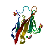
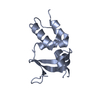
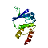
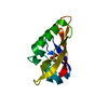
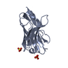
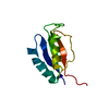
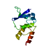
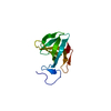
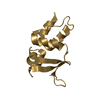
| Title | PDZ domain from rat Shank3 bound to the C terminus of GKAP | ||||||
 Components Components |
| ||||||
 Keywords Keywords |  PROTEIN BINDING / PDZ domain / peptide binding / post-synaptic density / C terminus PROTEIN BINDING / PDZ domain / peptide binding / post-synaptic density / C terminus | ||||||
| Function / homology |  Function and homology information Function and homology informationresponse to interleukin-17 / regulation of AMPA glutamate receptor clustering / guanylate kinase-associated protein clustering / striatal medium spiny neuron differentiation / synaptic receptor adaptor activity / maintenance of postsynaptic density structure / RET signaling / positive regulation of glutamate receptor signaling pathway / postsynaptic density assembly / embryonic epithelial tube formation ...response to interleukin-17 / regulation of AMPA glutamate receptor clustering / guanylate kinase-associated protein clustering / striatal medium spiny neuron differentiation / synaptic receptor adaptor activity / maintenance of postsynaptic density structure / RET signaling / positive regulation of glutamate receptor signaling pathway / postsynaptic density assembly / embryonic epithelial tube formation / postsynaptic specialization / Neurexins and neuroligins / positive regulation of synapse structural plasticity / signaling / negative regulation of actin filament bundle assembly / vocal learning / negative regulation of cell volume / positive regulation of long-term neuronal synaptic plasticity / structural constituent of postsynaptic density / regulation of grooming behavior / protein localization to synapse / NMDA glutamate receptor clustering / vocalization behavior / regulation of dendritic spine morphogenesis / regulation of behavioral fear response / neuron spine / AMPA glutamate receptor clustering / neural precursor cell proliferation / locomotion / dendritic spine morphogenesis / brain morphogenesis / aggresome assembly / regulation of long-term synaptic potentiation / positive regulation of AMPA receptor activity / long-term synaptic depression / regulation of postsynapse organization / ciliary membrane / exploration behavior / regulation of long-term synaptic depression / adult behavior / positive regulation of dendritic spine development / locomotory exploration behavior / postsynaptic density, intracellular component / social behavior / associative learning / positive regulation of excitatory postsynaptic potential / neuromuscular process controlling balance / glial cell proliferation / regulation of proteasomal protein catabolic process / synapse assembly / ionotropic glutamate receptor binding / positive regulation of synaptic transmission, glutamatergic / locomotory behavior / learning / long-term synaptic potentiation / positive regulation of long-term synaptic potentiation / G protein-coupled receptor binding / regulation of synaptic plasticity / modulation of chemical synaptic transmission / memory / SH3 domain binding / : / MAPK cascade / gene expression / actin binding / scaffold protein binding / postsynaptic membrane / dendritic spine / postsynaptic density / learning or memory / molecular adaptor activity / neuron projection / protein domain specific binding / synapse / glutamatergic synapse / protein-containing complex binding / zinc ion binding / identical protein binding / plasma membrane / cytoplasmSimilarity search - Function | ||||||
| Biological species |  Rattus norvegicus (Norway rat) Rattus norvegicus (Norway rat) | ||||||
| Method | X-RAY DIFFRACTION / SYNCHROTRON / MOLECULAR REPLACEMENT / Resolution: 1.55 Å | ||||||
 Authors Authors | Ponna, S.K. / Myllykoski, M. / Boeckers, T.M. / Kursula, P. | ||||||
 Citation Citation | Journal: J. Neurochem. / Year: 2018 Title: Structural basis for PDZ domain interactions in the post-synaptic density scaffolding protein Shank3. Authors: Ponna, S.K. / Ruskamo, S. / Myllykoski, M. / Keller, C. / Boeckers, T.M. / Kursula, P. | ||||||
| History |
|
- Structure visualization
| Structure viewer | Molecule: MolmilJmol/JSmol |
|---|
- Downloads & links
-Download
| PDBx/mmCIF format | 5ovc.cif.gz | 73.3 KB | Display | PDBx/mmCIF format |
|---|---|---|---|---|
| PDB format | pdb5ovc.ent.gz | 58.6 KB | Display | PDB format |
| PDBx/mmJSON format | 5ovc.json.gz | Tree view | PDBx/mmJSON format | |
| Others |  Other downloads Other downloads |
-Validation report
| Arichive directory | https://data.pdbj.org/pub/pdb/validation_reports/ov/5ovcftp://data.pdbj.org/pub/pdb/validation_reports/ov/5ovc | HTTPS FTP |
|---|
-Related structure data

-Links
 PDBj
PDBj
- Assembly
| Deposited unit | 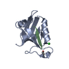
| ||||||||
|---|---|---|---|---|---|---|---|---|---|
| 1 |
| ||||||||
| Unit cell |
| ||||||||
| Components on special symmetry positions |
|
-Components
| #1: Protein | Mass: 10400.004 Da / Num. of mol.: 1 Source method: isolated from a genetically manipulated source Source: (gene. exp.) Rattus norvegicus (Norway rat) / Gene: Shank3, Prosap2 / Production host:  Escherichia coli (E. coli) / References: UniProt: Q9JLU4 Escherichia coli (E. coli) / References: UniProt: Q9JLU4 |
|---|---|
| #2: Protein/peptide | Mass: 743.829 Da / Num. of mol.: 1 / Source method: obtained synthetically / Source: (synth.) Rattus norvegicus (Norway rat) / References: UniProt: P97836*PLUS |
| #3: Chemical | ChemComp-CL / Chloride  Mass: 35.453 Da / Num. of mol.: 1 / Source method: obtained synthetically / Formula: Cl Mass: 35.453 Da / Num. of mol.: 1 / Source method: obtained synthetically / Formula: Cl |
| #4: Water | ChemComp-HOH / Water Mass: 18.015 Da / Num. of mol.: 131 / Source method: isolated from a natural source / Formula: H2O Mass: 18.015 Da / Num. of mol.: 131 / Source method: isolated from a natural source / Formula: H2O |
-Experimental details
-Experiment
| Experiment | Method: X-RAY DIFFRACTION / Number of used crystals: 1 |
|---|
- Sample preparation
| Crystal | Density Matthews: 2.44 Å3/Da / Density % sol: 49.54 % |
|---|---|
| Crystal grow | Temperature: 293 K / Method: vapor diffusion, sitting drop / Details: 1.6M sodium citrate pH 6.5 |
-Data collection
| Diffraction | Mean temperature: 100 K |
|---|---|
| Diffraction source | Source: SYNCHROTRON / Site: Diamond  / Beamline: I04 / Wavelength: 0.9795 Å / Beamline: I04 / Wavelength: 0.9795 Å |
| Detector | Type: DECTRIS PILATUS3 6M / Detector: PIXEL / Date: May 3, 2016 |
| Radiation | Protocol: SINGLE WAVELENGTH / Monochromatic (M) / Laue (L): M / Scattering type: x-ray |
| Radiation wavelength | Wavelength: 0.9795 Å / Relative weight: 1 |
| Reflection | Resolution: 1.55→50 Å / Num. obs: 30593 / % possible obs: 99.9 % / Redundancy: 6.7 % / CC1/2: 0.999 / Rrim(I) all: 0.126 / Net I/σ(I): 22.1 |
| Reflection shell | Resolution: 1.55→1.59 Å / Redundancy: 6 % / Num. unique obs: 2244 / CC1/2: 0.132 / Rrim(I) all: 3.628 / % possible all: 99.9 |
- Processing
| Software |
| |||||||||||||||||||||||||||||||||||||||||||||||||||||||||||||||||||||||||||||||||||||||||||||||||||||||||||||||||||||||||||||||||||||||||||||||||||||||||||||||||||||||||||||||
|---|---|---|---|---|---|---|---|---|---|---|---|---|---|---|---|---|---|---|---|---|---|---|---|---|---|---|---|---|---|---|---|---|---|---|---|---|---|---|---|---|---|---|---|---|---|---|---|---|---|---|---|---|---|---|---|---|---|---|---|---|---|---|---|---|---|---|---|---|---|---|---|---|---|---|---|---|---|---|---|---|---|---|---|---|---|---|---|---|---|---|---|---|---|---|---|---|---|---|---|---|---|---|---|---|---|---|---|---|---|---|---|---|---|---|---|---|---|---|---|---|---|---|---|---|---|---|---|---|---|---|---|---|---|---|---|---|---|---|---|---|---|---|---|---|---|---|---|---|---|---|---|---|---|---|---|---|---|---|---|---|---|---|---|---|---|---|---|---|---|---|---|---|---|---|---|---|
| Refinement | Method to determine structure: MOLECULAR REPLACEMENT / Resolution: 1.55→43.608 Å / SU ML: 0.23 / Cross valid method: FREE R-VALUE / σ(F): 1.32 / Phase error: 24.01
| |||||||||||||||||||||||||||||||||||||||||||||||||||||||||||||||||||||||||||||||||||||||||||||||||||||||||||||||||||||||||||||||||||||||||||||||||||||||||||||||||||||||||||||||
| Solvent computation | Shrinkage radii: 0.9 Å / VDW probe radii: 1.11 Å | |||||||||||||||||||||||||||||||||||||||||||||||||||||||||||||||||||||||||||||||||||||||||||||||||||||||||||||||||||||||||||||||||||||||||||||||||||||||||||||||||||||||||||||||
| Refinement step | Cycle: LAST / Resolution: 1.55→43.608 Å
| |||||||||||||||||||||||||||||||||||||||||||||||||||||||||||||||||||||||||||||||||||||||||||||||||||||||||||||||||||||||||||||||||||||||||||||||||||||||||||||||||||||||||||||||
| Refine LS restraints |
| |||||||||||||||||||||||||||||||||||||||||||||||||||||||||||||||||||||||||||||||||||||||||||||||||||||||||||||||||||||||||||||||||||||||||||||||||||||||||||||||||||||||||||||||
| LS refinement shell |
| |||||||||||||||||||||||||||||||||||||||||||||||||||||||||||||||||||||||||||||||||||||||||||||||||||||||||||||||||||||||||||||||||||||||||||||||||||||||||||||||||||||||||||||||
| Refinement TLS params. | Method: refined / Refine-ID: X-RAY DIFFRACTION
| |||||||||||||||||||||||||||||||||||||||||||||||||||||||||||||||||||||||||||||||||||||||||||||||||||||||||||||||||||||||||||||||||||||||||||||||||||||||||||||||||||||||||||||||
| Refinement TLS group |
|
