 Movie
Movie Controller

[English] 日本語
 Yorodumi
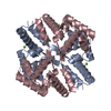
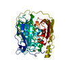
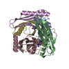
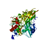
- PDB-1t1d: CRYSTAL STRUCTURE OF THE TETRAMERIZATION DOMAIN OF THE SHAKER POT... -
+ Open data

- Basic information
| Entry | Database: PDB / ID: 1t1d | ||||||
|---|---|---|---|---|---|---|---|
| Title | CRYSTAL STRUCTURE OF THE TETRAMERIZATION DOMAIN OF THE SHAKER POTASSIUM CHANNEL | ||||||
 Components Components | PROTEIN (POTASSIUM CHANNEL KV1.1) | ||||||
 Keywords Keywords |  MEMBRANE PROTEIN / POTASSIUM CHANNELS / TETRAMERIZATION DOMAIN / APLYSIA KV1.1 / PROTON TRANSPORT MEMBRANE PROTEIN / POTASSIUM CHANNELS / TETRAMERIZATION DOMAIN / APLYSIA KV1.1 / PROTON TRANSPORT | ||||||
| Function / homology |  Function and homology information Function and homology informationmonoatomic ion-gated channel activity / voltage-gated potassium channel activity / voltage-gated potassium channel complex / protein homooligomerizationSimilarity search - Function | ||||||
| Biological species |  Aplysia californica (California sea hare) Aplysia californica (California sea hare) | ||||||
| Method | X-RAY DIFFRACTION / SYNCHROTRON / MOLECULAR REPLACEMENT / Resolution: 1.51 Å | ||||||
 Authors Authors | Kreusch, A. / Pfaffinger, P.J. / Stevens, C.F. / Choe, S. | ||||||
 Citation Citation | Journal: Nat.Struct.Biol. / Year: 1999 Title: Zn2+-binding and molecular determinants of tetramerization in voltage-gated K+ channels. Authors: Bixby, K.A. / Nanao, M.H. / Shen, N.V. / Kreusch, A. / Bellamy, H. / Pfaffinger, P.J. / Choe, S. | ||||||
| History |
|
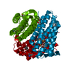
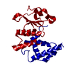
- Structure visualization
| Structure viewer | Molecule: MolmilJmol/JSmol |
|---|
- Downloads & links
-Download
| PDBx/mmCIF format | 1t1d.cif.gz | 34.7 KB | Display | PDBx/mmCIF format |
|---|---|---|---|---|
| PDB format | pdb1t1d.ent.gz | 23.9 KB | Display | PDB format |
| PDBx/mmJSON format | 1t1d.json.gz | Tree view | PDBx/mmJSON format | |
| Others |  Other downloads Other downloads |
-Validation report
| Arichive directory | https://data.pdbj.org/pub/pdb/validation_reports/t1/1t1dftp://data.pdbj.org/pub/pdb/validation_reports/t1/1t1d | HTTPS FTP |
|---|
-Related structure data
| Related structure data | 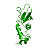 3kvtC  1a68S S: Starting model for refinement C: citing same article ( |
|---|---|
| Similar structure data |

-Links
 PDBj
PDBj


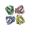
- Assembly
| Deposited unit | 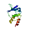
| ||||||||
|---|---|---|---|---|---|---|---|---|---|
| 1 | 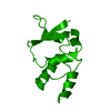
| ||||||||
| Unit cell |
|
-Components
| #1: Protein | Mass: 12097.453 Da / Num. of mol.: 1 / Fragment: TETRAMERIZATION DOMAIN Source method: isolated from a genetically manipulated source Source: (gene. exp.) Aplysia californica (California sea hare)Strain: BL21 (DE3) / Cell line: CENTRAL NERVOUS SYSTEM / Cellular location: CYTOPLASM / Plasmid: PET20B / Species (production host): Escherichia coli / Cellular location (production host): CYTOPLASM / Production host:  Escherichia coli BL21(DE3) (bacteria) / Strain (production host): BL21 (DE3) / References: UniProt: Q16968 Escherichia coli BL21(DE3) (bacteria) / Strain (production host): BL21 (DE3) / References: UniProt: Q16968 |
|---|---|
| #2: Water | ChemComp-HOH / Water Mass: 18.015 Da / Num. of mol.: 108 / Source method: isolated from a natural source / Formula: H2O Mass: 18.015 Da / Num. of mol.: 108 / Source method: isolated from a natural source / Formula: H2O |
-Experimental details
-Experiment
| Experiment | Method: X-RAY DIFFRACTION / Number of used crystals: 1 |
|---|
- Sample preparation
| Crystal | Density Matthews: 2.7 Å3/Da / Density % sol: 55 % | ||||||||||||||||||||||||||||||||||||||||||||||||||||||||||||||||||||||
|---|---|---|---|---|---|---|---|---|---|---|---|---|---|---|---|---|---|---|---|---|---|---|---|---|---|---|---|---|---|---|---|---|---|---|---|---|---|---|---|---|---|---|---|---|---|---|---|---|---|---|---|---|---|---|---|---|---|---|---|---|---|---|---|---|---|---|---|---|---|---|---|
| Crystal grow | pH: 7.5 Details: 24% ISOPROPANOL, .2M MGCL2, .1 M HEPES PH 7.5, 1MM DTT | ||||||||||||||||||||||||||||||||||||||||||||||||||||||||||||||||||||||
| Crystal | *PLUS | ||||||||||||||||||||||||||||||||||||||||||||||||||||||||||||||||||||||
| Crystal grow | *PLUS Method: vapor diffusion | ||||||||||||||||||||||||||||||||||||||||||||||||||||||||||||||||||||||
| Components of the solutions | *PLUS
|
-Data collection
| Diffraction | Mean temperature: 100 K |
|---|---|
| Diffraction source | Source: SYNCHROTRON / Site: SSRL  / Beamline: BL7-1 / Wavelength: 1.08 / Beamline: BL7-1 / Wavelength: 1.08 |
| Detector | Type: MARRESEARCH / Detector: IMAGE PLATE / Date: Apr 15, 1998 / Details: PT-COATED-MIRROR |
| Radiation | Monochromator: SI(111) / Protocol: SINGLE WAVELENGTH / Monochromatic (M) / Laue (L): M / Scattering type: x-ray |
| Radiation wavelength | Wavelength: 1.08 Å / Relative weight: 1 |
| Reflection | Resolution: 1.51→25 Å / Num. obs: 26768 / % possible obs: 92 % / Observed criterion σ(I): -3 / Redundancy: 5.9 % / Biso Wilson estimate: 19.35 Å2 / Rsym value: 0.049 / Net I/σ(I): 10.2 |
| Reflection shell | Resolution: 1.51→1.54 Å / Mean I/σ(I) obs: 3.5 / Rsym value: 0.16 / % possible all: 93.7 |
| Reflection | *PLUS Num. obs: 24631 / % possible obs: 93.7 % / Num. measured all: 135247 / Rmerge(I) obs: 0.066 |
- Processing
| Software |
| ||||||||||||||||||||||||||||||||||||||||||||||||||||||||||||||||||||||||||||||||||||
|---|---|---|---|---|---|---|---|---|---|---|---|---|---|---|---|---|---|---|---|---|---|---|---|---|---|---|---|---|---|---|---|---|---|---|---|---|---|---|---|---|---|---|---|---|---|---|---|---|---|---|---|---|---|---|---|---|---|---|---|---|---|---|---|---|---|---|---|---|---|---|---|---|---|---|---|---|---|---|---|---|---|---|---|---|---|
| Refinement | Method to determine structure: MOLECULAR REPLACEMENT Starting model: 1A68 Resolution: 1.51→19.8 Å / Cross valid method: THROUGHOUT / σ(F): 0
| ||||||||||||||||||||||||||||||||||||||||||||||||||||||||||||||||||||||||||||||||||||
| Refinement step | Cycle: LAST / Resolution: 1.51→19.8 Å
| ||||||||||||||||||||||||||||||||||||||||||||||||||||||||||||||||||||||||||||||||||||
| Refine LS restraints |
| ||||||||||||||||||||||||||||||||||||||||||||||||||||||||||||||||||||||||||||||||||||
| Software | *PLUS Name: REFMAC / Classification: refinement | ||||||||||||||||||||||||||||||||||||||||||||||||||||||||||||||||||||||||||||||||||||
| Refinement | *PLUS Lowest resolution: 19.8 Å / σ(F): 0 / % reflection Rfree: 5 % / Rfactor obs: 0.229 | ||||||||||||||||||||||||||||||||||||||||||||||||||||||||||||||||||||||||||||||||||||
| Solvent computation | *PLUS | ||||||||||||||||||||||||||||||||||||||||||||||||||||||||||||||||||||||||||||||||||||
| Displacement parameters | *PLUS | ||||||||||||||||||||||||||||||||||||||||||||||||||||||||||||||||||||||||||||||||||||
| Refine LS restraints | *PLUS
|
