 Movie
Movie Controller

[English] 日本語
 Yorodumi
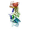
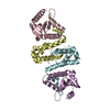
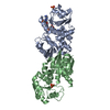
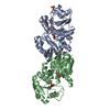
- PDB-5omn: GII.10 Vietnam 026 protruding domain in complex with Nanobody Nano-27 -
+ Open data

- Basic information
| Entry | Database: PDB / ID: 5omn | ||||||
|---|---|---|---|---|---|---|---|
| Title | GII.10 Vietnam 026 protruding domain in complex with Nanobody Nano-27 | ||||||
 Components Components |
| ||||||
 Keywords Keywords |  VIRAL PROTEIN / Norovirus / Protruding domain / capsid / VHH / Nanobody VIRAL PROTEIN / Norovirus / Protruding domain / capsid / VHH / Nanobody | ||||||
| Function / homology |  Function and homology information Function and homology informationPositive stranded ssRNA viruses / Nucleoplasmin-like/VP (viral coat and capsid proteins) / Positive stranded ssRNA viruses / Calicivirus coat protein C-terminal / Calicivirus coat protein C-terminal / Calicivirus coat protein / Calicivirus coat protein / Elongation Factor Tu (Ef-tu); domain 3 / Picornavirus/Calicivirus coat protein / Viral coat protein subunit ...Positive stranded ssRNA viruses / Nucleoplasmin-like/VP (viral coat and capsid proteins) / Positive stranded ssRNA viruses / Calicivirus coat protein C-terminal / Calicivirus coat protein C-terminal / Calicivirus coat protein / Calicivirus coat protein / Elongation Factor Tu (Ef-tu); domain 3 / Picornavirus/Calicivirus coat protein / Viral coat protein subunit / Beta Barrel / Mainly BetaSimilarity search - Domain/homology | ||||||
| Biological species |  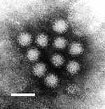 Norwalk virus Norwalk virus Vicugna pacos (alpaca) Vicugna pacos (alpaca) | ||||||
| Method | X-RAY DIFFRACTION / SYNCHROTRON / MOLECULAR REPLACEMENT / Resolution: 2.682 Å | ||||||
 Authors Authors | Koromyslova, A.D. / Hansman, G.S. | ||||||
 Citation Citation | Journal: PLoS Pathog. / Year: 2017 Title: Nanobodies targeting norovirus capsid reveal functional epitopes and potential mechanisms of neutralization. Authors: Koromyslova, A.D. / Hansman, G.S. | ||||||
| History |
|
- Structure visualization
| Structure viewer | Molecule: MolmilJmol/JSmol |
|---|
- Downloads & links
-Download
| PDBx/mmCIF format | 5omn.cif.gz | 94.5 KB | Display | PDBx/mmCIF format |
|---|---|---|---|---|
| PDB format | pdb5omn.ent.gz | 70.1 KB | Display | PDB format |
| PDBx/mmJSON format | 5omn.json.gz | Tree view | PDBx/mmJSON format | |
| Others |  Other downloads Other downloads |
-Validation report
| Arichive directory | https://data.pdbj.org/pub/pdb/validation_reports/om/5omnftp://data.pdbj.org/pub/pdb/validation_reports/om/5omn | HTTPS FTP |
|---|
-Related structure data
| Related structure data | 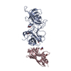 5o02C 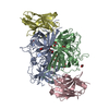 5o03C  5o04C 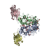 5o05C 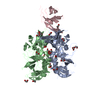 5ommC 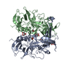 3onuS  4x7eS S: Starting model for refinement C: citing same article ( |
|---|---|
| Similar structure data |


-Links
 PDBj
PDBj


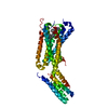
- Assembly
| Deposited unit | 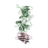
| ||||||||
|---|---|---|---|---|---|---|---|---|---|
| 1 |
| ||||||||
| Unit cell |
|
-Components
| #1: Protein | Capsid Mass: 34506.660 Da / Num. of mol.: 1 Source method: isolated from a genetically manipulated source Source: (gene. exp.) Norwalk virus / Plasmid: pMBP / Production host:  Escherichia coli BL21 (bacteria) / References: UniProt: Q5F4T5 Escherichia coli BL21 (bacteria) / References: UniProt: Q5F4T5 |
|---|---|
| #2: Antibody | Mass: 13392.866 Da / Num. of mol.: 1 Source method: isolated from a genetically manipulated source Source: (gene. exp.) Vicugna pacos (alpaca) / Plasmid: pHEN6C / Production host: Escherichia coli (E. coli) / Variant (production host): WK6 |
-Experimental details
-Experiment
| Experiment | Method: X-RAY DIFFRACTION / Number of used crystals: 1 |
|---|
- Sample preparation
| Crystal | Density Matthews: 6.22 Å3/Da / Density % sol: 80.22 % |
|---|---|
| Crystal grow | Temperature: 291 K / Method: vapor diffusion, hanging drop / Details: 0.1 M sodium acetate |
-Data collection
| Diffraction | Mean temperature: 100 K | ||||||||||||||||||||||||||||||||||||||||||||||||||||||||||||||||||||||||||||||||
|---|---|---|---|---|---|---|---|---|---|---|---|---|---|---|---|---|---|---|---|---|---|---|---|---|---|---|---|---|---|---|---|---|---|---|---|---|---|---|---|---|---|---|---|---|---|---|---|---|---|---|---|---|---|---|---|---|---|---|---|---|---|---|---|---|---|---|---|---|---|---|---|---|---|---|---|---|---|---|---|---|---|
| Diffraction source | Source: SYNCHROTRON / Site: ESRF  / Beamline: ID23-1 / Wavelength: 0.97319 Å / Beamline: ID23-1 / Wavelength: 0.97319 Å | ||||||||||||||||||||||||||||||||||||||||||||||||||||||||||||||||||||||||||||||||
| Detector | Type: DECTRIS PILATUS 6M-F / Detector: PIXEL / Date: Mar 8, 2014 | ||||||||||||||||||||||||||||||||||||||||||||||||||||||||||||||||||||||||||||||||
| Radiation | Protocol: SINGLE WAVELENGTH / Monochromatic (M) / Laue (L): M / Scattering type: x-ray | ||||||||||||||||||||||||||||||||||||||||||||||||||||||||||||||||||||||||||||||||
| Radiation wavelength | Wavelength: 0.97319 Å / Relative weight: 1 | ||||||||||||||||||||||||||||||||||||||||||||||||||||||||||||||||||||||||||||||||
| Reflection | Resolution: 2.68→48.338 Å / Num. obs: 33589 / % possible obs: 99.5 % / Observed criterion σ(I): -3 / Redundancy: 19.296 % / Biso Wilson estimate: 65.73 Å2 / CC1/2: 0.999 / Rmerge(I) obs: 0.15 / Rrim(I) all: 0.154 / Χ2: 1.156 / Net I/σ(I): 20.6 | ||||||||||||||||||||||||||||||||||||||||||||||||||||||||||||||||||||||||||||||||
| Reflection shell | Diffraction-ID: 1
|
- Processing
| Software |
| |||||||||||||||||||||||||||||||||||||||||||||||||||||||||||||||||||||||||||||||||||||||||||
|---|---|---|---|---|---|---|---|---|---|---|---|---|---|---|---|---|---|---|---|---|---|---|---|---|---|---|---|---|---|---|---|---|---|---|---|---|---|---|---|---|---|---|---|---|---|---|---|---|---|---|---|---|---|---|---|---|---|---|---|---|---|---|---|---|---|---|---|---|---|---|---|---|---|---|---|---|---|---|---|---|---|---|---|---|---|---|---|---|---|---|---|---|
| Refinement | Method to determine structure: MOLECULAR REPLACEMENT Starting model: 3ONU, 4X7E Resolution: 2.682→48.338 Å / SU ML: 0.43 / Cross valid method: FREE R-VALUE / σ(F): 1.36 / Phase error: 29.02
| |||||||||||||||||||||||||||||||||||||||||||||||||||||||||||||||||||||||||||||||||||||||||||
| Solvent computation | Shrinkage radii: 0.9 Å / VDW probe radii: 1.11 Å | |||||||||||||||||||||||||||||||||||||||||||||||||||||||||||||||||||||||||||||||||||||||||||
| Displacement parameters | Biso max: 145.44 Å2 / Biso mean: 68.2221 Å2 / Biso min: 21.44 Å2 | |||||||||||||||||||||||||||||||||||||||||||||||||||||||||||||||||||||||||||||||||||||||||||
| Refinement step | Cycle: final / Resolution: 2.682→48.338 Å
| |||||||||||||||||||||||||||||||||||||||||||||||||||||||||||||||||||||||||||||||||||||||||||
| Refine LS restraints |
| |||||||||||||||||||||||||||||||||||||||||||||||||||||||||||||||||||||||||||||||||||||||||||
| LS refinement shell | Refine-ID: X-RAY DIFFRACTION / Rfactor Rfree error: 0 / Total num. of bins used: 12
|
