 Movie
Movie Controller

[English] 日本語
 Yorodumi
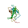
- PDB-5njo: Roll out the beta-barrel: structure and mechanism of Pac13, a uni... -
+ Open data

- Basic information
| Entry | Database: PDB / ID: 5njo | ||||||
|---|---|---|---|---|---|---|---|
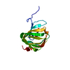
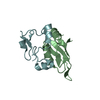
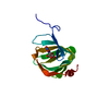
| Title | Roll out the beta-barrel: structure and mechanism of Pac13, a unique nucleoside dehydratase | ||||||
 Components Components | Putative cupin_2 domain-containing isomerase | ||||||
 Keywords Keywords |  STRUCTURAL PROTEIN / Pac13 Cupin Pacidamycin STRUCTURAL PROTEIN / Pac13 Cupin Pacidamycin | ||||||
| Function / homology | RmlC-like cupin domain superfamily / isomerase activity / RmlC-like jelly roll fold / Putative cupin_2 domain-containing isomerase Function and homology information Function and homology information | ||||||
| Biological species |  Streptomyces coeruleorubidus (bacteria) Streptomyces coeruleorubidus (bacteria) | ||||||
| Method | X-RAY DIFFRACTION / SYNCHROTRON / MOLECULAR REPLACEMENT / Resolution: 1.55 Å | ||||||
 Authors Authors | Michailidou, F. / Bent, A.F. / Naismith, J.H. / Goss, R.J.M. | ||||||
| Funding support |  United Kingdom, 1items United Kingdom, 1items
| ||||||
 Citation Citation | Journal: Angew. Chem. Int. Ed. Engl. / Year: 2017 Title: Pac13 is a Small, Monomeric Dehydratase that Mediates the Formation of the 3'-Deoxy Nucleoside of Pacidamycins. Authors: Michailidou, F. / Chung, C.W. / Brown, M.J.B. / Bent, A.F. / Naismith, J.H. / Leavens, W.J. / Lynn, S.M. / Sharma, S.V. / Goss, R.J.M. | ||||||
| History |
|
- Structure visualization
| Structure viewer | Molecule: MolmilJmol/JSmol |
|---|
- Downloads & links
-Download
| PDBx/mmCIF format | 5njo.cif.gz | 42.2 KB | Display | PDBx/mmCIF format |
|---|---|---|---|---|
| PDB format | pdb5njo.ent.gz | 28.6 KB | Display | PDB format |
| PDBx/mmJSON format | 5njo.json.gz | Tree view | PDBx/mmJSON format | |
| Others |  Other downloads Other downloads |
-Validation report
| Arichive directory | https://data.pdbj.org/pub/pdb/validation_reports/nj/5njoftp://data.pdbj.org/pub/pdb/validation_reports/nj/5njo | HTTPS FTP |
|---|
-Related structure data
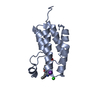
| Related structure data | 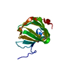 5njnSC  5oo4C  5oo5C 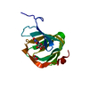 5oo8C 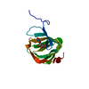 5oo9C 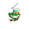 5ooaC S: Starting model for refinement C: citing same article ( |
|---|---|
| Similar structure data |


-Links
 PDBj
PDBj- Assembly
| Deposited unit | 
| ||||||||||||
|---|---|---|---|---|---|---|---|---|---|---|---|---|---|
| 1 |
| ||||||||||||
| Unit cell |
| ||||||||||||
| Components on special symmetry positions |
|
-Components
| #1: Protein | Mass: 13829.337 Da / Num. of mol.: 1 Source method: isolated from a genetically manipulated source Source: (gene. exp.) Streptomyces coeruleorubidus (bacteria)Gene: pac13, pacM / Production host: Escherichia coli BL21(DE3) (bacteria) / Variant (production host): RIPL / References: UniProt: E2EKP5 |
|---|---|
| #2: Water | ChemComp-HOH / Water Mass: 18.015 Da / Num. of mol.: 181 / Source method: isolated from a natural source / Formula: H2O Mass: 18.015 Da / Num. of mol.: 181 / Source method: isolated from a natural source / Formula: H2O |
-Experimental details
-Experiment
| Experiment | Method: X-RAY DIFFRACTION / Number of used crystals: 1 |
|---|
- Sample preparation
| Crystal | Density Matthews: 2.52 Å3/Da / Density % sol: 51.28 % |
|---|---|
| Crystal grow | Temperature: 293 K / Method: vapor diffusion, hanging drop / pH: 8.8 / Details: 0.1 M Tris pH 8.8, 20% w/v PEG8K, 0.2 M MgCl2 |
-Data collection
| Diffraction | Mean temperature: 100 K |
|---|---|
| Diffraction source | Source: SYNCHROTRON / Site: Diamond / Beamline: I03 / Wavelength: 0.9797 Å |
| Detector | Type: DECTRIS PILATUS 6M / Detector: PIXEL / Date: Feb 1, 2015 |
| Radiation | Protocol: SINGLE WAVELENGTH / Monochromatic (M) / Laue (L): M / Scattering type: x-ray |
| Radiation wavelength | Wavelength: 0.9797 Å / Relative weight: 1 |
| Reflection | Resolution: 1.55→57.58 Å / Num. obs: 20630 / % possible obs: 100 % / Redundancy: 10.9 % / Biso Wilson estimate: 18.63 Å2 / Rmerge(I) obs: 0.059 / Net I/σ(I): 23.5 |
| Reflection shell | Resolution: 1.55→1.61 Å / Redundancy: 10.6 % / Rmerge(I) obs: 0.776 / Num. unique obs: 2031 / % possible all: 99.9 |
- Processing
| Software |
| ||||||||||||||||||||||||||||||||||||||||||||||||||||||||||||||||||||||||||||||||||||||||||||||||||||||||||||||||||||||||||||||||||||||||||||||||||||||||||||||||||||||||||||||||||||||
|---|---|---|---|---|---|---|---|---|---|---|---|---|---|---|---|---|---|---|---|---|---|---|---|---|---|---|---|---|---|---|---|---|---|---|---|---|---|---|---|---|---|---|---|---|---|---|---|---|---|---|---|---|---|---|---|---|---|---|---|---|---|---|---|---|---|---|---|---|---|---|---|---|---|---|---|---|---|---|---|---|---|---|---|---|---|---|---|---|---|---|---|---|---|---|---|---|---|---|---|---|---|---|---|---|---|---|---|---|---|---|---|---|---|---|---|---|---|---|---|---|---|---|---|---|---|---|---|---|---|---|---|---|---|---|---|---|---|---|---|---|---|---|---|---|---|---|---|---|---|---|---|---|---|---|---|---|---|---|---|---|---|---|---|---|---|---|---|---|---|---|---|---|---|---|---|---|---|---|---|---|---|---|---|
| Refinement | Method to determine structure: MOLECULAR REPLACEMENT Starting model: 5NJN Resolution: 1.55→57.58 Å / Cor.coef. Fo:Fc: 0.965 / Cor.coef. Fo:Fc free: 0.956 / SU B: 1.393 / SU ML: 0.051 / Cross valid method: THROUGHOUT / ESU R: 0.079 / ESU R Free: 0.077 / Details: HYDROGENS HAVE BEEN ADDED IN THE RIDING POSITIONS
| ||||||||||||||||||||||||||||||||||||||||||||||||||||||||||||||||||||||||||||||||||||||||||||||||||||||||||||||||||||||||||||||||||||||||||||||||||||||||||||||||||||||||||||||||||||||
| Solvent computation | Ion probe radii: 0.8 Å / Shrinkage radii: 0.8 Å / VDW probe radii: 1.2 Å | ||||||||||||||||||||||||||||||||||||||||||||||||||||||||||||||||||||||||||||||||||||||||||||||||||||||||||||||||||||||||||||||||||||||||||||||||||||||||||||||||||||||||||||||||||||||
| Displacement parameters | Biso mean: 21.958 Å2
| ||||||||||||||||||||||||||||||||||||||||||||||||||||||||||||||||||||||||||||||||||||||||||||||||||||||||||||||||||||||||||||||||||||||||||||||||||||||||||||||||||||||||||||||||||||||
| Refinement step | Cycle: 1 / Resolution: 1.55→57.58 Å
| ||||||||||||||||||||||||||||||||||||||||||||||||||||||||||||||||||||||||||||||||||||||||||||||||||||||||||||||||||||||||||||||||||||||||||||||||||||||||||||||||||||||||||||||||||||||
| Refine LS restraints |
|