 Movie
Movie Controller

[English] 日本語
 Yorodumi
- PDB-1sz9: The RNA polymerase II CTD in mRNA processing: beta-turn recogniti... -
+ Open data

- Basic information
| Entry | Database: PDB / ID: 1sz9 | ||||||
|---|---|---|---|---|---|---|---|
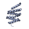
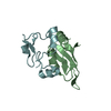
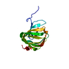
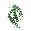
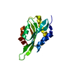
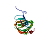
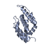
| Title | The RNA polymerase II CTD in mRNA processing: beta-turn recognition and beta-spiral model | ||||||
 Components Components | PCF11 protein | ||||||
 Keywords Keywords |  TRANSCRIPTION / Pcf11 / RNA polymerase II CTD interacting domain / arm repeats TRANSCRIPTION / Pcf11 / RNA polymerase II CTD interacting domain / arm repeats | ||||||
| Function / homology |  Function and homology information Function and homology informationtermination of RNA polymerase II transcription, poly(A)-coupled / termination of RNA polymerase II transcription, exosome-dependent / mRNA cleavage factor complex / termination of RNA polymerase II transcription / : / RNA polymerase II complex binding / mRNA binding / nucleus / cytosol / cytoplasmSimilarity search - Function | ||||||
| Biological species |  Saccharomyces cerevisiae (brewer's yeast) Saccharomyces cerevisiae (brewer's yeast) | ||||||
| Method | X-RAY DIFFRACTION / SYNCHROTRON / FOURIER SYNTHESIS / Resolution: 2.1 Å | ||||||
 Authors Authors | Meinhart, A. / Cramer, P. | ||||||
 Citation Citation | Journal: Nature / Year: 2004 Title: Recognition of RNA polymerase II carboxy-terminal domain by 3'-RNA-processing factors. Authors: Meinhart, A. / Cramer, P. | ||||||
| History |
|
- Structure visualization
| Structure viewer | Molecule: MolmilJmol/JSmol |
|---|
- Downloads & links
-Download
| PDBx/mmCIF format | 1sz9.cif.gz | 94.5 KB | Display | PDBx/mmCIF format |
|---|---|---|---|---|
| PDB format | pdb1sz9.ent.gz | 74.5 KB | Display | PDB format |
| PDBx/mmJSON format | 1sz9.json.gz | Tree view | PDBx/mmJSON format | |
| Others |  Other downloads Other downloads |
-Validation report
| Arichive directory | https://data.pdbj.org/pub/pdb/validation_reports/sz/1sz9ftp://data.pdbj.org/pub/pdb/validation_reports/sz/1sz9 | HTTPS FTP |
|---|
-Related structure data


-Links
 PDBj
PDBj
- Assembly
| Deposited unit | 
| ||||||||
|---|---|---|---|---|---|---|---|---|---|
| 1 | 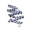
| ||||||||
| 2 | 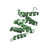
| ||||||||
| 3 | 
| ||||||||
| Unit cell |
|
-Components
| #1: Protein | Mass: 16624.145 Da / Num. of mol.: 3 / Fragment: CTD interacting domain of Pcf11 Source method: isolated from a genetically manipulated source Source: (gene. exp.) Saccharomyces cerevisiae (brewer's yeast)Gene: PCF11, YDR228C, YD9934.13C / Plasmid: pET28b / Production host:  Escherichia coli (E. coli) / Strain (production host): BL21(DE3)-RIL / References: UniProt: P39081 Escherichia coli (E. coli) / Strain (production host): BL21(DE3)-RIL / References: UniProt: P39081#2: Water | ChemComp-HOH / | Water Mass: 18.015 Da / Num. of mol.: 109 / Source method: isolated from a natural source / Formula: H2O Mass: 18.015 Da / Num. of mol.: 109 / Source method: isolated from a natural source / Formula: H2O |
|---|
-Experimental details
-Experiment
| Experiment | Method: X-RAY DIFFRACTION / Number of used crystals: 1 |
|---|
- Sample preparation
| Crystal | Density Matthews: 2.61 Å3/Da / Density % sol: 52.86 % |
|---|---|
| Crystal grow | Temperature: 298 K / pH: 7.5 Details: 25 % (v/v) ethylene glycol, 5 % (v/v) PEG 550, pH 7.5, VAPOR DIFFUSION, HANGING DROP, temperature 298.0K, pH 7.50 |
-Data collection
| Diffraction | Mean temperature: 100 K |
|---|---|
| Diffraction source | Source: SYNCHROTRON / Site: ESRF  / Beamline: ID29 / Wavelength: 0.9168 / Beamline: ID29 / Wavelength: 0.9168 |
| Detector | Type: ADSC QUANTUM 4 / Detector: CCD / Date: Sep 9, 2003 |
| Radiation | Protocol: MAD / Monochromatic (M) / Laue (L): M / Scattering type: x-ray |
| Radiation wavelength | Wavelength: 0.9168 Å / Relative weight: 1 |
| Reflection | Resolution: 2.1→20 Å / Num. obs: 29988 / % possible obs: 98 % / Observed criterion σ(I): 3 / Redundancy: 4.7 % / Rsym value: 0.065 / Net I/σ(I): 13.5 |
| Reflection shell | Resolution: 2.1→2.2 Å / Mean I/σ(I) obs: 4.5 / Rsym value: 0.307 / % possible all: 96.7 |
- Processing
| Software |
| ||||||||||||||||||||||||||||||||||||||||||||||||||||||||||||
|---|---|---|---|---|---|---|---|---|---|---|---|---|---|---|---|---|---|---|---|---|---|---|---|---|---|---|---|---|---|---|---|---|---|---|---|---|---|---|---|---|---|---|---|---|---|---|---|---|---|---|---|---|---|---|---|---|---|---|---|---|---|
| Refinement | Method to determine structure: FOURIER SYNTHESIS Starting model: ORIGINAL MODEL FROM MAD EXPERIMENT WAS USED FOR FURTHER REFINEMENT. R-FREE SET WAS KEPT CONSISTEND Resolution: 2.1→20 Å / σ(F): 3 / Stereochemistry target values: ENGH & HUBER
| ||||||||||||||||||||||||||||||||||||||||||||||||||||||||||||
| Refinement step | Cycle: LAST / Resolution: 2.1→20 Å
| ||||||||||||||||||||||||||||||||||||||||||||||||||||||||||||
| Refine LS restraints |
| ||||||||||||||||||||||||||||||||||||||||||||||||||||||||||||
| LS refinement shell | Resolution: 2.1→2.13 Å / Total num. of bins used: 28 /
|
