[English] 日本語
Yorodumi
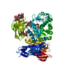
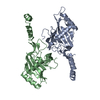
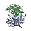
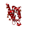
- PDB-6i2v: Pilotin from Vibrio vulnificus type 2 secretion system, EpsS. -

+
Open data


ID or keywords:

Loading...

-
Basic information

Entry
Database: PDB / ID: 6i2v
TitlePilotin from Vibrio vulnificus type 2 secretion system, EpsS.
ComponentsUncharacterized protein
KeywordsPROTEIN BINDING / Pilotin / T2SS / V.vulnificus / Outer-membrane
Function / homologyGMP Synthetase; Chain A, domain 3 - #250 / Type II secretion system (T2SS) pilotin, S protein / Type II secretion system (T2SS) pilotin, S protein / GMP Synthetase; Chain A, domain 3 / 2-Layer Sandwich / Alpha Beta / SULFITE ION / Uncharacterized protein
Function and homology information
Biological speciesVibrio vulnificus (bacteria)
MethodX-RAY DIFFRACTION / SYNCHROTRON / MOLECULAR REPLACEMENT / Resolution: 1.75 Å
AuthorsHoward, S.P. / Estrozi, L. / Bertrand, Q. / Contreras-Martel, C. / Strozen, T. / Job, V. / Martins, A. / Fenel, D. / Schoehn, G. / Dessen, A.
CitationJournal: PLoS Pathog / Year: 2019
Title: Structure and assembly of pilotin-dependent and -independent secretins of the type II secretion system.
Authors: S Peter Howard / Leandro F Estrozi / Quentin Bertrand / Carlos Contreras-Martel / Timothy Strozen / Viviana Job / Alexandre Martins / Daphna Fenel / Guy Schoehn / Andréa Dessen /
Abstract: The type II secretion system (T2SS) is a cell envelope-spanning macromolecular complex that is prevalent in Gram-negative bacterial species. It serves as the predominant virulence mechanism of many ...The type II secretion system (T2SS) is a cell envelope-spanning macromolecular complex that is prevalent in Gram-negative bacterial species. It serves as the predominant virulence mechanism of many bacteria including those of the emerging human pathogens Vibrio vulnificus and Aeromonas hydrophila. The system is composed of a core set of highly conserved proteins that assemble an inner membrane platform, a periplasmic pseudopilus and an outer membrane complex termed the secretin. Localization and assembly of secretins in the outer membrane requires recognition of secretin monomers by two different partner systems: an inner membrane accessory complex or a highly sequence-diverse outer membrane lipoprotein, termed the pilotin. In this study, we addressed the question of differential secretin assembly mechanisms by using cryo-electron microscopy to determine the structures of the secretins from A. hydrophila (pilotin-independent ExeD) and V. vulnificus (pilotin-dependent EpsD). These structures, at approximately 3.5 Å resolution, reveal pentadecameric stoichiometries and C-terminal regions that carry a signature motif in the case of a pilotin-dependent assembly mechanism. We solved the crystal structure of the V. vulnificus EpsS pilotin and confirmed the importance of the signature motif for pilotin-dependent secretin assembly by performing modelling with the C-terminus of EpsD. We also show that secretin assembly is essential for membrane integrity and toxin secretion in V. vulnificus and establish that EpsD requires the coordinated activity of both the accessory complex EpsAB and the pilotin EpsS for full assembly and T2SS function. In contrast, mutation of the region of the S-domain that is normally the site of pilotin interactions has little effect on assembly or function of the ExeD secretin. Since secretins are essential outer membrane channels present in a variety of secretion systems, these results provide a structural and functional basis for understanding the key assembly steps for different members of this vast pore-forming family of proteins.
History
DepositionNov 2, 2018Deposition site: PDBE / Processing site: PDBE
Revision 1.0Apr 10, 2019Provider: repository / Type: Initial release
Revision 1.1Aug 28, 2019Group: Data collection / Database references / Category: citation / citation_author / reflns
Item: _citation.country / _citation.journal_abbrev ..._citation.country / _citation.journal_abbrev / _citation.journal_id_CSD / _citation.journal_id_ISSN / _citation.journal_volume / _citation.page_first / _citation.page_last / _citation.pdbx_database_id_DOI / _citation.pdbx_database_id_PubMed / _citation.title / _citation.year / _citation_author.identifier_ORCID / _citation_author.name / _reflns.pdbx_CC_half / _reflns.pdbx_Rsym_value
Revision 1.2Jan 24, 2024Group: Data collection / Database references / Refinement description
Category: chem_comp_atom / chem_comp_bond ...chem_comp_atom / chem_comp_bond / database_2 / pdbx_initial_refinement_model
Item: _database_2.pdbx_DOI / _database_2.pdbx_database_accession

-
Structure visualization

Structure viewerMolecule:
MolmilJmol/JSmol

Downloads & links

-
Assembly

Deposited unit
A: Uncharacterized protein
hetero molecules


Theoretical massNumber of molelcules
Total (without water)14,39014
Polymers12,5141
Non-polymers1,87613
Water1,71195
1
A: Uncharacterized protein
hetero molecules

A: Uncharacterized protein
hetero molecules

A: Uncharacterized protein
hetero molecules

A: Uncharacterized protein
hetero molecules


Theoretical massNumber of molelcules
Total (without water)57,56156
Polymers50,0574
Non-polymers7,50452
Water724
TypeNameSymmetry operationNumber
identity operation1_555x,y,z1
crystal symmetry operation2_555-x,-y,z1
crystal symmetry operation3_556-x,y,-z+11
crystal symmetry operation4_556x,-y,-z+11
Buried area12530 Å2
ΔGint-249 kcal/mol
Surface area23220 Å2
MethodPISA
Unit cell
Length a, b, c (Å)35.830, 83.210, 90.790
Angle α, β, γ (deg.)90.00, 90.00, 90.00
Int Tables number23
Space group name H-MI222
Components on special symmetry positions
IDModelComponents
11A-212-

SO4

21A-388-

HOH

-
Components

-
Protein , 1 types, 1 molecules A

#1: Protein Uncharacterized protein


Mass: 12514.362 Da / Num. of mol.: 1
Source method: isolated from a genetically manipulated source
Source: (gene. exp.) Vibrio vulnificus (bacteria) / Gene: CRN52_08940, CRN59_22290, FORC17_1995, FORC36_0970 / Plasmid: p4-MBP / Production host: Escherichia coli (E. coli) / Strain (production host): Bl21 / References: UniProt: A0A1V8MYF4

-
Non-polymers , 5 types, 108 molecules

#2: Chemical
ChemComp-1PE / PENTAETHYLENE GLYCOL / PEG400 / Polyethylene glycol


Mass: 238.278 Da / Num. of mol.: 5 / Source method: obtained synthetically / Formula: C10H22O6 / Comment: precipitant*YM
#3: Chemical ChemComp-EDO / 1,2-ETHANEDIOL / ETHYLENE GLYCOL / Ethylene glycol


Mass: 62.068 Da / Num. of mol.: 2 / Source method: obtained synthetically / Formula: C2H6O2
#4: Chemical ChemComp-SO3 / SULFITE ION / Sulfite


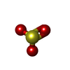
Mass: 80.063 Da / Num. of mol.: 1 / Source method: obtained synthetically / Formula: SO3
#5: Chemical
ChemComp-SO4 / SULFATE ION / Sulfate


Mass: 96.063 Da / Num. of mol.: 5 / Source method: obtained synthetically / Formula: SO4
#6: Water ChemComp-HOH / water / Water


Mass: 18.015 Da / Num. of mol.: 95 / Source method: isolated from a natural source / Formula: H2O

-
Experimental details

-
Experiment

ExperimentMethod: X-RAY DIFFRACTION / Number of used crystals: 1

-
Sample preparation

CrystalDensity Matthews: 2.7 Å3/Da / Density % sol: 54.51 %
Crystal growTemperature: 293.15 K / Method: vapor diffusion
Details: 0.1M tris Hcl pH 7.5 1.9M Amonium Sulfate 2.5% (v/v) PEG400

-
Data collection

DiffractionMean temperature: 100 K / Serial crystal experiment: N
Diffraction sourceSource: SYNCHROTRON / Site: ESRF / Beamline: MASSIF-3 / Wavelength: 0.9677 Å
DetectorType: DECTRIS EIGER X 4M / Detector: PIXEL / Date: Apr 23, 2018
RadiationProtocol: SINGLE WAVELENGTH / Monochromatic (M) / Laue (L): M / Scattering type: x-ray
Radiation wavelengthWavelength: 0.9677 Å / Relative weight: 1
ReflectionResolution: 1.75→45.39 Å / Num. obs: 14037 / % possible obs: 99.3 % / Redundancy: 16 % / Biso Wilson estimate: 28.85 Å2 / CC1/2: 0.999 / Rsym value: 0.13 / Net I/σ(I): 14.14
Reflection shellResolution: 1.75→1.85 Å / Mean I/σ(I) obs: 2.12 / CC1/2: 0.88 / Rsym value: 1.15 / % possible all: 95.8

-
Processing

Software
NameVersionClassification
REFMAC5.8.0232refinement
XDSdata reduction
XDSdata scaling
Arcimboldophasing
RefinementMethod to determine structure: MOLECULAR REPLACEMENT
Starting model: 4FTF
Resolution: 1.75→45.39 Å / Cor.coef. Fo:Fc: 0.964 / Cor.coef. Fo:Fc free: 0.939 / SU B: 5.128 / SU ML: 0.081 / Cross valid method: THROUGHOUT / ESU R: 0.12 / ESU R Free: 0.123 / Details: HYDROGENS HAVE BEEN ADDED IN THE RIDING POSITIONS
RfactorNum. reflection% reflectionSelection details
Rfree0.22787 1822 13 %RANDOM
Rwork0.17904 ---
obs0.18546 12215 99.38 %-
Solvent computationIon probe radii: 0.8 Å / Shrinkage radii: 0.8 Å / VDW probe radii: 1.2 Å
Displacement parametersBiso mean: 25.385 Å2
Baniso -1Baniso -2Baniso -3
1--1.34 Å2-0 Å20 Å2
2---1.03 Å2-0 Å2
3---2.37 Å2
Refinement stepCycle: 1 / Resolution: 1.75→45.39 Å
ProteinNucleic acidLigandSolventTotal
Num. atoms843 0 69 95 1007
Refine LS restraints
Refine-IDTypeDev idealDev ideal targetNumber
X-RAY DIFFRACTIONr_bond_refined_d0.0130.013968
X-RAY DIFFRACTIONr_bond_other_d0.0010.017941
X-RAY DIFFRACTIONr_angle_refined_deg1.3971.671298
X-RAY DIFFRACTIONr_angle_other_deg1.3041.6272199
X-RAY DIFFRACTIONr_dihedral_angle_1_deg6.2275127
X-RAY DIFFRACTIONr_dihedral_angle_2_deg33.97922.72744
X-RAY DIFFRACTIONr_dihedral_angle_3_deg13.74915189
X-RAY DIFFRACTIONr_dihedral_angle_4_deg7.273157
X-RAY DIFFRACTIONr_chiral_restr0.0790.2128
X-RAY DIFFRACTIONr_gen_planes_refined0.0120.021016
X-RAY DIFFRACTIONr_gen_planes_other0.0030.02165
X-RAY DIFFRACTIONr_nbd_refined
X-RAY DIFFRACTIONr_nbd_other
X-RAY DIFFRACTIONr_nbtor_refined
X-RAY DIFFRACTIONr_nbtor_other
X-RAY DIFFRACTIONr_xyhbond_nbd_refined
X-RAY DIFFRACTIONr_xyhbond_nbd_other
X-RAY DIFFRACTIONr_metal_ion_refined
X-RAY DIFFRACTIONr_metal_ion_other
X-RAY DIFFRACTIONr_symmetry_vdw_refined
X-RAY DIFFRACTIONr_symmetry_vdw_other
X-RAY DIFFRACTIONr_symmetry_hbond_refined
X-RAY DIFFRACTIONr_symmetry_hbond_other
X-RAY DIFFRACTIONr_symmetry_metal_ion_refined
X-RAY DIFFRACTIONr_symmetry_metal_ion_other
X-RAY DIFFRACTIONr_mcbond_it1.1491.498458
X-RAY DIFFRACTIONr_mcbond_other0.9871.487456
X-RAY DIFFRACTIONr_mcangle_it1.352.221574
X-RAY DIFFRACTIONr_mcangle_other1.352.223575
X-RAY DIFFRACTIONr_scbond_it2.6282.028510
X-RAY DIFFRACTIONr_scbond_other2.4231.908491
X-RAY DIFFRACTIONr_scangle_it
X-RAY DIFFRACTIONr_scangle_other2.7562.666686
X-RAY DIFFRACTIONr_long_range_B_refined6.14919.7741016
X-RAY DIFFRACTIONr_long_range_B_other6.09919.1131000
X-RAY DIFFRACTIONr_rigid_bond_restr
X-RAY DIFFRACTIONr_sphericity_free
X-RAY DIFFRACTIONr_sphericity_bonded
LS refinement shellResolution: 1.75→1.796 Å / Total num. of bins used: 20
RfactorNum. reflection% reflection
Rfree0.345 97 -
Rwork0.352 853 -
obs--92.23 %
Refinement TLS params.

Method: refined / Refine-ID: X-RAY DIFFRACTION

IDL112)L122)L132)L222)L232)L332)S11 (Å °)S12 (Å °)S13 (Å °)S21 (Å °)S22 (Å °)S23 (Å °)S31 (Å °)S32 (Å °)S33 (Å °)T112)T122)T132)T222)T232)T332)Origin x (Å)Origin y (Å)Origin z (Å)
19.57190.8948-0.307816.7233-3.338813.4667-0.16391.22950.4437-0.9538-0.0103-0.361-0.22770.26220.17420.18650.05260.04020.21930.05370.172220.566.559410.7469
24.4115-0.2777-0.92034.8853-4.867511.79540.04330.18250.2048-0.1256-0.252-0.3519-0.17510.41030.20870.07870.0141-0.00080.05550.00960.133917.94964.793121.4183
310.3694.1269-6.26951.746-2.25155.46830.1444-0.42330.03970.0517-0.0393-0.0154-0.07760.4583-0.10510.0712-0.0058-0.02740.161-0.03580.079212.07822.963435.884
48.1938-7.08175.94769.56-0.356311.69220.08740.16690.4892-0.22210.2343-0.6867-0.27270.5757-0.32170.112-0.01240.06690.1001-0.0070.19144.396910.675340.6694
51.847-1.1458-2.83562.72783.979811.2581-0.17010.0422-0.02440.22870.06860.15890.5383-0.30270.10150.0917-0.0066-0.00480.05850.02560.09446.44643.079130.1679
610.25061.78-10.21753.2425-2.24216.7878-0.12730.1959-0.1558-0.10170.05230.00710.2864-0.24570.07490.09480.0074-0.03090.05220.01710.11436.96286.267124.3532
71.28850.7562-0.49031.2731-1.88593.2680.07870.02020.15850.110.0510.1334-0.1418-0.0782-0.12960.10990.01930.06840.05090.03690.0686-6.259912.287938.657
810.34224.3819-2.02125.7624-4.10254.7947-0.0697-0.19950.05910.3802-0.0078-0.1602-0.23090.05050.07750.09590.01030.01720.06850.00830.07130.724320.379231.9625
914.98412.1891-1.99548.9522-3.29965.0166-0.0714-0.78530.34860.60560.07760.0383-0.1841-0.1901-0.00620.10040.00820.00760.10910.02280.076710.63119.59230.9882
101.4620.84150.71362.17510.36534.36690.0487-0.0553-0.09310.0056-0.0749-0.28880.1680.02310.02610.06910.00810.00570.060.04020.126217.272416.180624.4368
113.86651.2835-2.59691.6409-0.78221.7498-0.03040.20940.02750.04880.04870.08260.026-0.1464-0.01820.0751-0.0039-0.00790.07250.03640.0825-1.19188.15628.7079
123.09082.0491-0.71471.6323-0.92221.2169-0.04150.11930.2968-0.0350.03790.1956-0.0785-0.00290.00360.07650.01390.00970.04970.04590.12915.99619.591421.8918
Refinement TLS group
IDRefine-IDRefine TLS-IDAuth asym-IDAuth seq-ID
1X-RAY DIFFRACTION1A5 - 10
2X-RAY DIFFRACTION2A11 - 21
3X-RAY DIFFRACTION3A22 - 29
4X-RAY DIFFRACTION4A30 - 34
5X-RAY DIFFRACTION5A35 - 42
6X-RAY DIFFRACTION6A43 - 50
7X-RAY DIFFRACTION7A51 - 62
8X-RAY DIFFRACTION8A63 - 70
9X-RAY DIFFRACTION9A71 - 75
10X-RAY DIFFRACTION10A76 - 87
11X-RAY DIFFRACTION11A88 - 101
12X-RAY DIFFRACTION12A102 - 114

+
About Yorodumi

-
News

-
Feb 9, 2022. New format data for meta-information of EMDB entries

New format data for meta-information of EMDB entries

  • Version 3 of the EMDB header file is now the official format.
  • The previous official version 1.9 will be removed from the archive.

Related info.:EMDB header

External links:wwPDB to switch to version 3 of the EMDB data model

-
Aug 12, 2020. Covid-19 info

Covid-19 info

URL: https://pdbj.org/emnavi/covid19.php

New page: Covid-19 featured information page in EM Navigator.

Related info.:Covid-19 info / Mar 5, 2020. Novel coronavirus structure data

+
Mar 5, 2020. Novel coronavirus structure data

Novel coronavirus structure data

Related info.:Yorodumi Speices / Aug 12, 2020. Covid-19 info

External links:COVID-19 featured content - PDBj / Molecule of the Month (242):Coronavirus Proteases

+
Jan 31, 2019. EMDB accession codes are about to change! (news from PDBe EMDB page)

EMDB accession codes are about to change! (news from PDBe EMDB page)

  • The allocation of 4 digits for EMDB accession codes will soon come to an end. Whilst these codes will remain in use, new EMDB accession codes will include an additional digit and will expand incrementally as the available range of codes is exhausted. The current 4-digit format prefixed with “EMD-” (i.e. EMD-XXXX) will advance to a 5-digit format (i.e. EMD-XXXXX), and so on. It is currently estimated that the 4-digit codes will be depleted around Spring 2019, at which point the 5-digit format will come into force.
  • The EM Navigator/Yorodumi systems omit the EMD- prefix.

Related info.:Q: What is EMD? / ID/Accession-code notation in Yorodumi/EM Navigator

External links:EMDB Accession Codes are Changing Soon! / Contact to PDBj

+
Jul 12, 2017. Major update of PDB

Major update of PDB

  • wwPDB released updated PDB data conforming to the new PDBx/mmCIF dictionary.
  • This is a major update changing the version number from 4 to 5, and with Remediation, in which all the entries are updated.
  • In this update, many items about electron microscopy experimental information are reorganized (e.g. em_software).
  • Now, EM Navigator and Yorodumi are based on the updated data.

External links:wwPDB Remediation / Enriched Model Files Conforming to OneDep Data Standards Now Available in the PDB FTP Archive

-
Yorodumi

Thousand views of thousand structures

  • Yorodumi is a browser for structure data from EMDB, PDB, SASBDB, etc.
  • This page is also the successor to EM Navigator detail page, and also detail information page/front-end page for Omokage search.
  • The word "yorodu" (or yorozu) is an old Japanese word meaning "ten thousand". "mi" (miru) is to see.

Related info.:EMDB / PDB / SASBDB / Comparison of 3 databanks / Yorodumi Search / Aug 31, 2016. New EM Navigator & Yorodumi / Yorodumi Papers / Jmol/JSmol / Function and homology information / Changes in new EM Navigator and Yorodumi

Read more