 Movie
Movie Controller

[English] 日本語
 Yorodumi
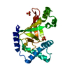
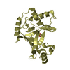
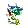
- PDB-5nbg: Structure of the cytoplasmic domain I of OutF in the D. dadantii ... -
+ Open data

- Basic information
| Entry | Database: PDB / ID: 5nbg | ||||||
|---|---|---|---|---|---|---|---|
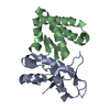
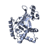
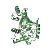
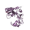
| Title | Structure of the cytoplasmic domain I of OutF in the D. dadantii type II secretion system | ||||||
 Components Components | General secretion pathway protein F | ||||||
 Keywords Keywords |  MEMBRANE PROTEIN / inner membrane protein MEMBRANE PROTEIN / inner membrane protein | ||||||
| Function / homology |  Function and homology informationprotein secretion by the type II secretion system / type II protein secretion system complex / protein processing / membrane => GO:0016020 / metal ion binding / plasma membrane Function and homology informationprotein secretion by the type II secretion system / type II protein secretion system complex / protein processing / membrane => GO:0016020 / metal ion binding / plasma membraneSimilarity search - Function | ||||||
| Biological species |  Dickeya dadantii (bacteria) Dickeya dadantii (bacteria) | ||||||
| Method | X-RAY DIFFRACTION / SYNCHROTRON / MOLECULAR REPLACEMENT / Resolution: 2.15 Å | ||||||
 Authors Authors | Zhang, H. / Gu, S. | ||||||
 Citation Citation | Journal: To Be Published Title: Structure of the first cytoplasmic domain of OutF and assembly of the inner-membrane platform proteins of the D. dadantii type II secretion system Authors: Zhang, H. / Gu, S. | ||||||
| History |
|
- Structure visualization
| Structure viewer | Molecule: MolmilJmol/JSmol |
|---|
- Downloads & links
-Download
| PDBx/mmCIF format | 5nbg.cif.gz | 52 KB | Display | PDBx/mmCIF format |
|---|---|---|---|---|
| PDB format | pdb5nbg.ent.gz | 38.1 KB | Display | PDB format |
| PDBx/mmJSON format | 5nbg.json.gz | Tree view | PDBx/mmJSON format | |
| Others |  Other downloads Other downloads |
-Validation report
| Arichive directory | https://data.pdbj.org/pub/pdb/validation_reports/nb/5nbgftp://data.pdbj.org/pub/pdb/validation_reports/nb/5nbg | HTTPS FTP |
|---|
-Related structure data
| Related structure data |  3c1qS S: Starting model for refinement |
|---|---|
| Similar structure data |


-Links
 PDBj
PDBj
- Assembly
| Deposited unit | 
| ||||||||
|---|---|---|---|---|---|---|---|---|---|
| 1 |
| ||||||||
| Unit cell |
|
-Components
| #1: Protein | Mass: 11667.410 Da / Num. of mol.: 2 / Fragment: UNP residues 65-172 Source method: isolated from a genetically manipulated source Source: (gene. exp.) Dickeya dadantii (strain 3937) (bacteria)Strain: 3937 / Gene: outF, Dda3937_02417 / Production host: Escherichia coli (E. coli) / References: UniProt: E0SM39#2: Water | ChemComp-HOH / | Water Mass: 18.015 Da / Num. of mol.: 53 / Source method: isolated from a natural source / Formula: H2O Mass: 18.015 Da / Num. of mol.: 53 / Source method: isolated from a natural source / Formula: H2O |
|---|
-Experimental details
-Experiment
| Experiment | Method: X-RAY DIFFRACTION / Number of used crystals: 1 |
|---|
- Sample preparation
| Crystal | Density Matthews: 3.5 Å3/Da / Density % sol: 64.86 % |
|---|---|
| Crystal grow | Temperature: 291 K / Method: vapor diffusion, hanging drop Details: 100mM sodium cacodyl ate pH 6.5, 200mM Li2SO4 and 30% PEG-400 |
-Data collection
| Diffraction | Mean temperature: 100 K |
|---|---|
| Diffraction source | Source: SYNCHROTRON / Site: ESRF  / Beamline: ID23-1 / Wavelength: 0.972422 Å / Beamline: ID23-1 / Wavelength: 0.972422 Å |
| Detector | Type: DECTRIS PILATUS 6M-F / Detector: PIXEL / Date: Oct 30, 2015 |
| Radiation | Protocol: SINGLE WAVELENGTH / Monochromatic (M) / Laue (L): M / Scattering type: x-ray |
| Radiation wavelength | Wavelength: 0.972422 Å / Relative weight: 1 |
| Reflection | Resolution: 2.15→41.44 Å / Num. obs: 18300 / % possible obs: 99.15 % / Redundancy: 5.5 % / CC1/2: 0.999 / Rmerge(I) obs: 0.062 / Rpim(I) all: 0.037 / Net I/σ(I): 1.35 |
| Reflection shell | Resolution: 2.15→2.23 Å / Redundancy: 5.9 % / Rmerge(I) obs: 0.024 / Num. unique obs: 1764 / CC1/2: 0.668 / Rpim(I) all: 0.016 / % possible all: 99.8 |
- Processing
| Software |
| ||||||||||||||||||||||||||||||||||||||||||||||||||||||||||||||||||||||||||||||||||||||||||||||||||||||||||||||||||||||||||||||||||||||||||||||||||||||||||||||||||||||||||||||||||||||
|---|---|---|---|---|---|---|---|---|---|---|---|---|---|---|---|---|---|---|---|---|---|---|---|---|---|---|---|---|---|---|---|---|---|---|---|---|---|---|---|---|---|---|---|---|---|---|---|---|---|---|---|---|---|---|---|---|---|---|---|---|---|---|---|---|---|---|---|---|---|---|---|---|---|---|---|---|---|---|---|---|---|---|---|---|---|---|---|---|---|---|---|---|---|---|---|---|---|---|---|---|---|---|---|---|---|---|---|---|---|---|---|---|---|---|---|---|---|---|---|---|---|---|---|---|---|---|---|---|---|---|---|---|---|---|---|---|---|---|---|---|---|---|---|---|---|---|---|---|---|---|---|---|---|---|---|---|---|---|---|---|---|---|---|---|---|---|---|---|---|---|---|---|---|---|---|---|---|---|---|---|---|---|---|
| Refinement | Method to determine structure: MOLECULAR REPLACEMENT Starting model: 3C1Q Resolution: 2.15→82.15 Å / Cor.coef. Fo:Fc: 0.953 / Cor.coef. Fo:Fc free: 0.924 / SU B: 5.98 / SU ML: 0.15 / Cross valid method: THROUGHOUT / ESU R: 0.192 / ESU R Free: 0.187 / Stereochemistry target values: MAXIMUM LIKELIHOOD / Details: HYDROGENS HAVE BEEN ADDED IN THE RIDING POSITIONS
| ||||||||||||||||||||||||||||||||||||||||||||||||||||||||||||||||||||||||||||||||||||||||||||||||||||||||||||||||||||||||||||||||||||||||||||||||||||||||||||||||||||||||||||||||||||||
| Solvent computation | Ion probe radii: 0.8 Å / Shrinkage radii: 0.8 Å / VDW probe radii: 1.2 Å / Solvent model: MASK | ||||||||||||||||||||||||||||||||||||||||||||||||||||||||||||||||||||||||||||||||||||||||||||||||||||||||||||||||||||||||||||||||||||||||||||||||||||||||||||||||||||||||||||||||||||||
| Displacement parameters | Biso mean: 51.982 Å2
| ||||||||||||||||||||||||||||||||||||||||||||||||||||||||||||||||||||||||||||||||||||||||||||||||||||||||||||||||||||||||||||||||||||||||||||||||||||||||||||||||||||||||||||||||||||||
| Refinement step | Cycle: 1 / Resolution: 2.15→82.15 Å
| ||||||||||||||||||||||||||||||||||||||||||||||||||||||||||||||||||||||||||||||||||||||||||||||||||||||||||||||||||||||||||||||||||||||||||||||||||||||||||||||||||||||||||||||||||||||
| Refine LS restraints |
|