 Movie
Movie Controller

[English] 日本語
 Yorodumi
- PDB-3vtg: High choriolytic enzyme 1 (HCE-1), a hatching enzyme zinc-proteas... -
+ Open data

- Basic information
| Entry | Database: PDB / ID: 3vtg | ||||||
|---|---|---|---|---|---|---|---|
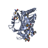
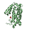
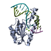
| Title | High choriolytic enzyme 1 (HCE-1), a hatching enzyme zinc-protease from Oryzias latipes (Medaka fish) | ||||||
 Components Components | High choriolytic enzyme 1 | ||||||
 Keywords Keywords |  HYDROLASE / hatching enzyme / Structural Genomics / NPPSFA / National Project on Protein Structural and Functional Analyses / astacin family / zinc protease HYDROLASE / hatching enzyme / Structural Genomics / NPPSFA / National Project on Protein Structural and Functional Analyses / astacin family / zinc protease | ||||||
| Function / homology |  Function and homology informationchoriolysin H / transport vesicle / metalloendopeptidase activity / zinc ion binding Function and homology informationchoriolysin H / transport vesicle / metalloendopeptidase activity / zinc ion bindingSimilarity search - Function | ||||||
| Biological species |  Oryzias latipes (Japanese medaka) Oryzias latipes (Japanese medaka) | ||||||
| Method | X-RAY DIFFRACTION / SYNCHROTRON / MOLECULAR REPLACEMENT / Resolution: 1.34 Å | ||||||
 Authors Authors | Kudo, N. / Yasumasu, S. / Iuchi, I. / Tanokura, M. | ||||||
 Citation Citation | Journal: To be Published Title: Crystal Structure of High choriolytic enzyme 1 (HCE-1), a hatching enzyme from Oryzias latipes (Medaka fish) Authors: Kudo, N. / Yasumasu, S. / Iuchi, I. / Tanokura, M. | ||||||
| History |
|
- Structure visualization
| Structure viewer | Molecule: MolmilJmol/JSmol |
|---|
- Downloads & links
-Download
| PDBx/mmCIF format | 3vtg.cif.gz | 55.1 KB | Display | PDBx/mmCIF format |
|---|---|---|---|---|
| PDB format | pdb3vtg.ent.gz | 42 KB | Display | PDB format |
| PDBx/mmJSON format | 3vtg.json.gz | Tree view | PDBx/mmJSON format | |
| Others |  Other downloads Other downloads |
-Validation report
| Arichive directory | https://data.pdbj.org/pub/pdb/validation_reports/vt/3vtgftp://data.pdbj.org/pub/pdb/validation_reports/vt/3vtg | HTTPS FTP |
|---|
-Related structure data
| Similar structure data |
|---|


-Links
 PDBj
PDBj


- Assembly
| Deposited unit | 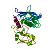
| |||||||||||||||
|---|---|---|---|---|---|---|---|---|---|---|---|---|---|---|---|---|
| 1 |
| |||||||||||||||
| Unit cell |
| |||||||||||||||
| Components on special symmetry positions |
|
-Components
| #1: Protein | Mass: 22716.467 Da / Num. of mol.: 1 / Source method: isolated from a natural source / Source: (natural) Oryzias latipes (Japanese medaka) / References: UniProt: P31580, choriolysin H |
|---|---|
| #2: Chemical | ChemComp-ZN /   Mass: 65.409 Da / Num. of mol.: 1 / Source method: obtained synthetically / Formula: Zn Mass: 65.409 Da / Num. of mol.: 1 / Source method: obtained synthetically / Formula: Zn |
| #3: Water | ChemComp-HOH / Water Mass: 18.015 Da / Num. of mol.: 294 / Source method: isolated from a natural source / Formula: H2O Mass: 18.015 Da / Num. of mol.: 294 / Source method: isolated from a natural source / Formula: H2O |
-Experimental details
-Experiment
| Experiment | Method: X-RAY DIFFRACTION / Number of used crystals: 1 |
|---|
- Sample preparation
| Crystal | Density Matthews: 2.2 Å3/Da / Density % sol: 44.08 % |
|---|---|
| Crystal grow | Temperature: 277 K / Method: vapor diffusion, hanging drop / Details: VAPOR DIFFUSION, HANGING DROP, temperature 277K |
-Data collection
| Diffraction | Mean temperature: 100 K |
|---|---|
| Diffraction source | Source: SYNCHROTRON / Site: SPring-8  / Beamline: BL41XU / Beamline: BL41XU |
| Detector | Type: MAR CCD 130 mm / Detector: CCD |
| Radiation | Protocol: SINGLE WAVELENGTH / Monochromatic (M) / Laue (L): M / Scattering type: x-ray |
| Radiation wavelength | Relative weight: 1 |
| Reflection | Resolution: 1.34→30 Å / Num. obs: 44714 / % possible obs: 99.3 % / Observed criterion σ(F): 0 / Observed criterion σ(I): 0 |
- Processing
| Software |
| |||||||||||||||||||||||||||||||||||||||||||||
|---|---|---|---|---|---|---|---|---|---|---|---|---|---|---|---|---|---|---|---|---|---|---|---|---|---|---|---|---|---|---|---|---|---|---|---|---|---|---|---|---|---|---|---|---|---|---|
| Refinement | Method to determine structure: MOLECULAR REPLACEMENT / Resolution: 1.34→20 Å / Cor.coef. Fo:Fc: 0.957 / Cor.coef. Fo:Fc free: 0.949 / Occupancy max: 1 / Occupancy min: 0.5 / SU B: 0.787 / SU ML: 0.034 / Cross valid method: THROUGHOUT / σ(F): 0 / ESU R: 0.059 / ESU R Free: 0.059 / Stereochemistry target values: MAXIMUM LIKELIHOOD Details: HYDROGENS HAVE BEEN USED IF PRESENT IN THE INPUT U VALUES: REFINED INDIVIDUALLY
| |||||||||||||||||||||||||||||||||||||||||||||
| Solvent computation | Ion probe radii: 0.8 Å / Shrinkage radii: 0.8 Å / VDW probe radii: 1.2 Å / Solvent model: MASK | |||||||||||||||||||||||||||||||||||||||||||||
| Displacement parameters | Biso max: 45.18 Å2 / Biso mean: 11.8342 Å2 / Biso min: 7.1 Å2
| |||||||||||||||||||||||||||||||||||||||||||||
| Refinement step | Cycle: LAST / Resolution: 1.34→20 Å
| |||||||||||||||||||||||||||||||||||||||||||||
| Refine LS restraints |
| |||||||||||||||||||||||||||||||||||||||||||||
| LS refinement shell | Resolution: 1.34→1.375 Å / Total num. of bins used: 20
|
