 Movie
Movie Controller


+ Open data

- Basic information
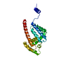
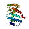
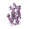
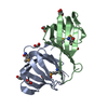
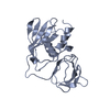
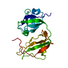
| Entry | Database: PDB / ID: 5mjo | |||||||||
|---|---|---|---|---|---|---|---|---|---|---|
| Title | Structure of Psb29 at 1.55A | |||||||||
 Components Components | Protein Thf1 | |||||||||
 Keywords Keywords |  PHOTOSYNTHESIS / photosystem II FtsH PHOTOSYNTHESIS / photosystem II FtsH | |||||||||
| Function / homology | Protein Thf1 / Thylakoid formation protein / photosystem II assembly / : / IODIDE ION / Protein Thf1 Function and homology information Function and homology information | |||||||||
| Biological species |   Thermosynechococcus elongatus (bacteria) Thermosynechococcus elongatus (bacteria) | |||||||||
| Method | X-RAY DIFFRACTION / SYNCHROTRON / SAD / Resolution: 1.55 Å | |||||||||
 Authors Authors | Murray, J.W. / Kozlo, A. | |||||||||
| Funding support |  United Kingdom, 2items United Kingdom, 2items
| |||||||||
 Citation Citation | Journal: Philos. Trans. R. Soc. Lond., B, Biol. Sci. / Year: 2017 Title: Structure of Psb29/Thf1 and its association with the FtsH protease complex involved in photosystem II repair in cyanobacteria. Authors: Bec Kova, M. / Yu, J. / Krynicka, V. / Kozlo, A. / Shao, S. / Konik, P. / Komenda, J. / Murray, J.W. / Nixon, P.J. | |||||||||
| History |
|
- Structure visualization
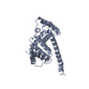
| Structure viewer | Molecule: MolmilJmol/JSmol |
|---|
- Downloads & links
-Download
| PDBx/mmCIF format | 5mjo.cif.gz | 97.1 KB | Display | PDBx/mmCIF format |
|---|---|---|---|---|
| PDB format | pdb5mjo.ent.gz | 72.8 KB | Display | PDB format |
| PDBx/mmJSON format | 5mjo.json.gz | Tree view | PDBx/mmJSON format | |
| Others |  Other downloads Other downloads |
-Validation report
| Arichive directory | https://data.pdbj.org/pub/pdb/validation_reports/mj/5mjoftp://data.pdbj.org/pub/pdb/validation_reports/mj/5mjo | HTTPS FTP |
|---|
-Related structure data


-Links
 PDBj
PDBj


- Assembly
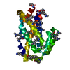
| Deposited unit | 
| ||||||||
|---|---|---|---|---|---|---|---|---|---|
| 1 |
| ||||||||
| Unit cell |
|
-Components
| #1: Protein | Mass: 27268.848 Da / Num. of mol.: 1 Source method: isolated from a genetically manipulated source Source: (gene. exp.) Thermosynechococcus elongatus (bacteria)Gene: thf1, tlr1134 / Plasmid: pRSETa modified / Production host: Escherichia coli (E. coli) / Strain (production host): KRX / References: UniProt: Q8DJT8 | ||
|---|---|---|---|
| #2: Chemical | ChemComp-HG / Mercury (element)  Mass: 200.590 Da / Num. of mol.: 1 / Source method: obtained synthetically / Formula: Hg Mass: 200.590 Da / Num. of mol.: 1 / Source method: obtained synthetically / Formula: Hg | ||
| #3: Chemical | Iodide  Mass: 126.904 Da / Num. of mol.: 2 / Source method: obtained synthetically / Formula: I Mass: 126.904 Da / Num. of mol.: 2 / Source method: obtained synthetically / Formula: I#4: Water | ChemComp-HOH / | Water Mass: 18.015 Da / Num. of mol.: 110 / Source method: isolated from a natural source / Formula: H2O Mass: 18.015 Da / Num. of mol.: 110 / Source method: isolated from a natural source / Formula: H2O |
-Experimental details
-Experiment
| Experiment | Method: X-RAY DIFFRACTION / Number of used crystals: 1 |
|---|
- Sample preparation
| Crystal | Density Matthews: 1.95 Å3/Da / Density % sol: 37 % |
|---|---|
| Crystal grow | Temperature: 290 K / Method: vapor diffusion, hanging drop / pH: 5 / Details: 0.1 M Bicine, pH 9.0, 20% w/v PEG 6000 |
-Data collection
| Diffraction | Mean temperature: 100 K |
|---|---|
| Diffraction source | Source: SYNCHROTRON / Site: Diamond / Beamline: I04-1 / Wavelength: 0.9173 Å |
| Detector | Type: DECTRIS PILATUS 2M / Detector: PIXEL / Date: Mar 3, 2012 |
| Radiation | Protocol: SINGLE WAVELENGTH / Monochromatic (M) / Laue (L): M / Scattering type: x-ray |
| Radiation wavelength | Wavelength: 0.9173 Å / Relative weight: 1 |
| Reflection | Resolution: 1.55→46.16 Å / Num. obs: 23219 / % possible obs: 99 % / Observed criterion σ(F): 0 / Observed criterion σ(I): 0 / Redundancy: 16.9 % / Biso Wilson estimate: 15.949 Å2 / Rmerge(I) obs: 0.114 / Net I/av σ(I): 18.3675 / Net I/σ(I): 5.2976 |
| Reflection shell | Resolution: 1.55→1.59 Å / Redundancy: 16.68 % / Rmerge(I) obs: 1.098 / Mean I/σ(I) obs: 0.65 / % possible all: 99.17 |
- Processing
| Software |
| ||||||||||||||||||||||||||||||||||||||||||||||||||||||||||||||||||||||||||||||||||||||||||||||||||||||||||||||||||||||||||||||||||||||||||||||||||||||||||||||||||||||||||||||||||||||
|---|---|---|---|---|---|---|---|---|---|---|---|---|---|---|---|---|---|---|---|---|---|---|---|---|---|---|---|---|---|---|---|---|---|---|---|---|---|---|---|---|---|---|---|---|---|---|---|---|---|---|---|---|---|---|---|---|---|---|---|---|---|---|---|---|---|---|---|---|---|---|---|---|---|---|---|---|---|---|---|---|---|---|---|---|---|---|---|---|---|---|---|---|---|---|---|---|---|---|---|---|---|---|---|---|---|---|---|---|---|---|---|---|---|---|---|---|---|---|---|---|---|---|---|---|---|---|---|---|---|---|---|---|---|---|---|---|---|---|---|---|---|---|---|---|---|---|---|---|---|---|---|---|---|---|---|---|---|---|---|---|---|---|---|---|---|---|---|---|---|---|---|---|---|---|---|---|---|---|---|---|---|---|---|
| Refinement | Method to determine structure: SAD / Resolution: 1.55→46.16 Å / Cor.coef. Fo:Fc: 0.972 / Cor.coef. Fo:Fc free: 0.954 / SU B: 3.561 / SU ML: 0.057 / Cross valid method: THROUGHOUT / ESU R: 0.1 / ESU R Free: 0.083 / Details: HYDROGENS HAVE BEEN ADDED IN THE RIDING POSITIONS
| ||||||||||||||||||||||||||||||||||||||||||||||||||||||||||||||||||||||||||||||||||||||||||||||||||||||||||||||||||||||||||||||||||||||||||||||||||||||||||||||||||||||||||||||||||||||
| Solvent computation | Ion probe radii: 0.8 Å / Shrinkage radii: 0.8 Å / VDW probe radii: 1.2 Å | ||||||||||||||||||||||||||||||||||||||||||||||||||||||||||||||||||||||||||||||||||||||||||||||||||||||||||||||||||||||||||||||||||||||||||||||||||||||||||||||||||||||||||||||||||||||
| Displacement parameters | Biso mean: 18.479 Å2
| ||||||||||||||||||||||||||||||||||||||||||||||||||||||||||||||||||||||||||||||||||||||||||||||||||||||||||||||||||||||||||||||||||||||||||||||||||||||||||||||||||||||||||||||||||||||
| Refinement step | Cycle: 1 / Resolution: 1.55→46.16 Å
| ||||||||||||||||||||||||||||||||||||||||||||||||||||||||||||||||||||||||||||||||||||||||||||||||||||||||||||||||||||||||||||||||||||||||||||||||||||||||||||||||||||||||||||||||||||||
| Refine LS restraints |
|