 Movie
Movie Controller


+ Open data

- Basic information
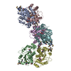
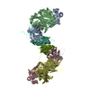
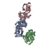
| Entry | Database: PDB / ID: 5mtv | ||||||
|---|---|---|---|---|---|---|---|
| Title | Active structure of EHD4 complexed with ATP-gamma-S | ||||||
 Components Components | EH domain-containing protein 4 | ||||||
 Keywords Keywords |  ENDOCYTOSIS / dynamin-like / membrane protein / auto-inhibition / activation ENDOCYTOSIS / dynamin-like / membrane protein / auto-inhibition / activation | ||||||
| Function / homology |  Function and homology informationpinocytosis / endocytic recycling / regulation of endocytosis / cilium assembly / endocytic vesicle / protein localization to plasma membrane / protein homooligomerization / cellular response to growth factor stimulus / endocytosis / recycling endosome membrane ...pinocytosis / endocytic recycling / regulation of endocytosis / cilium assembly / endocytic vesicle / protein localization to plasma membrane / protein homooligomerization / cellular response to growth factor stimulus / endocytosis / recycling endosome membrane / positive regulation of peptidyl-tyrosine phosphorylation / early endosome membrane / early endosome / calcium ion binding / GTP binding / perinuclear region of cytoplasm / endoplasmic reticulum / ATP binding / plasma membrane / cytoplasm Function and homology informationpinocytosis / endocytic recycling / regulation of endocytosis / cilium assembly / endocytic vesicle / protein localization to plasma membrane / protein homooligomerization / cellular response to growth factor stimulus / endocytosis / recycling endosome membrane ...pinocytosis / endocytic recycling / regulation of endocytosis / cilium assembly / endocytic vesicle / protein localization to plasma membrane / protein homooligomerization / cellular response to growth factor stimulus / endocytosis / recycling endosome membrane / positive regulation of peptidyl-tyrosine phosphorylation / early endosome membrane / early endosome / calcium ion binding / GTP binding / perinuclear region of cytoplasm / endoplasmic reticulum / ATP binding / plasma membrane / cytoplasmSimilarity search - Function | ||||||
| Biological species |  Mus musculus (house mouse) Mus musculus (house mouse) | ||||||
| Method | X-RAY DIFFRACTION / SYNCHROTRON / MOLECULAR REPLACEMENT / Resolution: 2.79 Å | ||||||
 Authors Authors | Melo, A.A. / Daumke, O. | ||||||
| Funding support |  Germany, 1items Germany, 1items
| ||||||
 Citation Citation | Journal: Proc. Natl. Acad. Sci. U.S.A. / Year: 2017 Title: Structural insights into the activation mechanism of dynamin-like EHD ATPases. Authors: Melo, A.A. / Hegde, B.G. / Shah, C. / Larsson, E. / Isas, J.M. / Kunz, S. / Lundmark, R. / Langen, R. / Daumke, O. | ||||||
| History |
|
- Structure visualization
| Structure viewer | Molecule: MolmilJmol/JSmol |
|---|
- Downloads & links
-Download
| PDBx/mmCIF format | 5mtv.cif.gz | 241.9 KB | Display | PDBx/mmCIF format |
|---|---|---|---|---|
| PDB format | pdb5mtv.ent.gz | 198.3 KB | Display | PDB format |
| PDBx/mmJSON format | 5mtv.json.gz | Tree view | PDBx/mmJSON format | |
| Others |  Other downloads Other downloads |
-Validation report
| Arichive directory | https://data.pdbj.org/pub/pdb/validation_reports/mt/5mtvftp://data.pdbj.org/pub/pdb/validation_reports/mt/5mtv | HTTPS FTP |
|---|
-Related structure data
| Related structure data | 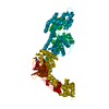 5mvfC  4cidS S: Starting model for refinement C: citing same article ( |
|---|---|
| Similar structure data |


-Links
 PDBj
PDBj
- Assembly
| Deposited unit | 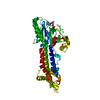
| ||||||||
|---|---|---|---|---|---|---|---|---|---|
| 1 | 
| ||||||||
| Unit cell |
| ||||||||
| Components on special symmetry positions |
|
-Components
| #1: Protein | Mass: 59488.074 Da / Num. of mol.: 1 Source method: isolated from a genetically manipulated source Source: (gene. exp.) Mus musculus (house mouse) / Gene: Ehd4, Past2 / Production host:  Escherichia coli (E. coli) / References: UniProt: Q9EQP2 Escherichia coli (E. coli) / References: UniProt: Q9EQP2 |
|---|---|
| #2: Chemical | ChemComp-AGS /   Mass: 523.247 Da / Num. of mol.: 1 / Source method: obtained synthetically / Formula: C10H16N5O12P3S / Comment: ATP-gamma-S, energy-carrying molecule analogue*YM Mass: 523.247 Da / Num. of mol.: 1 / Source method: obtained synthetically / Formula: C10H16N5O12P3S / Comment: ATP-gamma-S, energy-carrying molecule analogue*YM |
| #3: Chemical | ChemComp-MG /   Mass: 24.305 Da / Num. of mol.: 1 / Source method: obtained synthetically / Formula: Mg Mass: 24.305 Da / Num. of mol.: 1 / Source method: obtained synthetically / Formula: Mg |
| #4: Water | ChemComp-HOH / Water Mass: 18.015 Da / Num. of mol.: 21 / Source method: isolated from a natural source / Formula: H2O Mass: 18.015 Da / Num. of mol.: 21 / Source method: isolated from a natural source / Formula: H2O |
-Experimental details
-Experiment
| Experiment | Method: X-RAY DIFFRACTION / Number of used crystals: 1 |
|---|
- Sample preparation
| Crystal | Density Matthews: 3.5 Å3/Da / Density % sol: 64.86 % |
|---|---|
| Crystal grow | Temperature: 293 K / Method: vapor diffusion, hanging drop / pH: 7.5 Details: Sodium polyacrylate 5100, magnesium chloride, HEPES |
-Data collection
| Diffraction | Mean temperature: 100 K |
|---|---|
| Diffraction source | Source: SYNCHROTRON / Site: BESSY / Beamline: 14.1 / Wavelength: 0.918409 Å |
| Detector | Type: DECTRIS PILATUS 6M / Detector: PIXEL / Date: Jul 19, 2015 / Details: DOUBLE MIRROR |
| Radiation | Monochromator: double Si-111 crystal / Protocol: SINGLE WAVELENGTH / Monochromatic (M) / Laue (L): M / Scattering type: x-ray |
| Radiation wavelength | Wavelength: 0.918409 Å / Relative weight: 1 |
| Reflection | Resolution: 2.79→48.5 Å / Num. obs: 21733 / % possible obs: 99.8 % / Observed criterion σ(F): -3 / Redundancy: 7.2 % / Biso Wilson estimate: 64.3 Å2 / CC1/2: 0.999 / Rmerge(I) obs: 0.095 / Rpim(I) all: 0.035 / Rsym value: 0.088 / Net I/σ(I): 19.04 |
| Reflection shell | Resolution: 2.79→2.96 Å / Redundancy: 7.65 % / Rmerge(I) obs: 0.91 / Mean I/σ(I) obs: 2.15 / Num. unique obs: 3412 / CC1/2: 0.69 / % possible all: 99.2 |
- Processing
| Software |
| ||||||||||||||||||||||||||||||||||||||||||||||||||||||||||||||||||||||||||||||||||||||||||||||||||||
|---|---|---|---|---|---|---|---|---|---|---|---|---|---|---|---|---|---|---|---|---|---|---|---|---|---|---|---|---|---|---|---|---|---|---|---|---|---|---|---|---|---|---|---|---|---|---|---|---|---|---|---|---|---|---|---|---|---|---|---|---|---|---|---|---|---|---|---|---|---|---|---|---|---|---|---|---|---|---|---|---|---|---|---|---|---|---|---|---|---|---|---|---|---|---|---|---|---|---|---|---|---|
| Refinement | Method to determine structure: MOLECULAR REPLACEMENT Starting model: 4CID Resolution: 2.79→48.5 Å / SU ML: 0.45 / Cross valid method: NONE / σ(F): 1.38 / Phase error: 25.07
| ||||||||||||||||||||||||||||||||||||||||||||||||||||||||||||||||||||||||||||||||||||||||||||||||||||
| Solvent computation | Shrinkage radii: 0.9 Å / VDW probe radii: 1.11 Å | ||||||||||||||||||||||||||||||||||||||||||||||||||||||||||||||||||||||||||||||||||||||||||||||||||||
| Displacement parameters | Biso mean: 82.316 Å2 | ||||||||||||||||||||||||||||||||||||||||||||||||||||||||||||||||||||||||||||||||||||||||||||||||||||
| Refinement step | Cycle: LAST / Resolution: 2.79→48.5 Å
| ||||||||||||||||||||||||||||||||||||||||||||||||||||||||||||||||||||||||||||||||||||||||||||||||||||
| Refine LS restraints |
| ||||||||||||||||||||||||||||||||||||||||||||||||||||||||||||||||||||||||||||||||||||||||||||||||||||
| LS refinement shell |
| ||||||||||||||||||||||||||||||||||||||||||||||||||||||||||||||||||||||||||||||||||||||||||||||||||||
| Refinement TLS params. | Method: refined / Refine-ID: X-RAY DIFFRACTION
| ||||||||||||||||||||||||||||||||||||||||||||||||||||||||||||||||||||||||||||||||||||||||||||||||||||
| Refinement TLS group |
|
