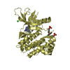
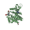
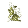
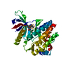
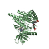
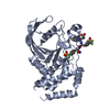
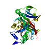
[English] 日本語
Yorodumi
- PDB-3zep: Crystal Structure of JAK3 Kinase Domain in Complex with a Pyrrolo... -

+
Open data


ID or keywords:

Loading...

-
Basic information

Entry
Database: PDB / ID: 3zep
TitleCrystal Structure of JAK3 Kinase Domain in Complex with a Pyrrolopyrazine-2-phenyl Ether Inhibitor
ComponentsTYROSINE-PROTEIN KINASE JAK3
KeywordsTRANSFERASE / STAT5 / STAT6 / INTERLEUKIN-2 / COMMON-GAMMA CHAIN / ATP SITE KINASE INHIBITOR / CANCER / SCID / SEVERE COMBINED IMMUNODEFICIENCY
Function / homology
Function and homology information


negative regulation of dendritic cell cytokine production / negative regulation of FasL production / response to interleukin-9 / response to interleukin-2 / response to interleukin-15 / response to interleukin-4 / negative regulation of T-helper 1 cell differentiation / negative regulation of T cell activation / Interleukin-9 signaling / Interleukin-21 signaling ...negative regulation of dendritic cell cytokine production / negative regulation of FasL production / response to interleukin-9 / response to interleukin-2 / response to interleukin-15 / response to interleukin-4 / negative regulation of T-helper 1 cell differentiation / negative regulation of T cell activation / Interleukin-9 signaling / Interleukin-21 signaling / interleukin-9-mediated signaling pathway / interleukin-4-mediated signaling pathway / interleukin-2-mediated signaling pathway / regulation of T cell apoptotic process / negative regulation of interleukin-12 production / interleukin-15-mediated signaling pathway / tyrosine phosphorylation of STAT protein / negative regulation of thymocyte apoptotic process / Interleukin-15 signaling / Interleukin-2 signaling / regulation of receptor signaling pathway via JAK-STAT / growth hormone receptor binding / extrinsic component of plasma membrane / Signaling by ALK / negative regulation of interleukin-10 production / Interleukin-20 family signaling / enzyme-linked receptor protein signaling pathway / T cell homeostasis / cell surface receptor signaling pathway via JAK-STAT / growth hormone receptor signaling pathway via JAK-STAT / Interleukin receptor SHC signaling / extrinsic component of cytoplasmic side of plasma membrane / Interleukin-7 signaling / B cell differentiation / non-specific protein-tyrosine kinase / non-membrane spanning protein tyrosine kinase activity / cytokine-mediated signaling pathway / peptidyl-tyrosine phosphorylation / RAF/MAP kinase cascade / regulation of apoptotic process / protein phosphatase binding / protein tyrosine kinase activity / Interleukin-4 and Interleukin-13 signaling / adaptive immune response / Potential therapeutics for SARS / cell differentiation / cytoskeleton / endosome / intracellular signal transduction / protein phosphorylation / innate immune response / ATP binding / plasma membrane / cytosol
Similarity search - Function
Tyrosine-protein kinase, non-receptor Jak3 / Tyrosine-protein kinase, non-receptor Jak/Tyk2 / JAK, FERM F2 lobe domain / FERM F1 lobe ubiquitin-like domain / JAK1-3/TYK2, pleckstrin homology-like domain / Jak1 pleckstrin homology-like domain / FERM F2 acyl-CoA binding protein-like domain / FERM F1 ubiquitin-like domain / FERM domain / FERM domain profile. ...Tyrosine-protein kinase, non-receptor Jak3 / Tyrosine-protein kinase, non-receptor Jak/Tyk2 / JAK, FERM F2 lobe domain / FERM F1 lobe ubiquitin-like domain / JAK1-3/TYK2, pleckstrin homology-like domain / Jak1 pleckstrin homology-like domain / FERM F2 acyl-CoA binding protein-like domain / FERM F1 ubiquitin-like domain / FERM domain / FERM domain profile. / Band 4.1 domain / Band 4.1 homologues / Src homology 2 domains / SH2 domain / SH2 domain superfamily / Tyrosine-protein kinase, catalytic domain / Tyrosine kinase, catalytic domain / Tyrosine protein kinases specific active-site signature. / PH-like domain superfamily / Tyrosine-protein kinase, active site / Protein tyrosine and serine/threonine kinase / Serine-threonine/tyrosine-protein kinase, catalytic domain / Transferase(Phosphotransferase) domain 1 / Transferase(Phosphotransferase); domain 1 / Phosphorylase Kinase; domain 1 / Phosphorylase Kinase; domain 1 / Protein kinase, ATP binding site / Protein kinases ATP-binding region signature. / Protein kinase domain profile. / Protein kinase domain / Protein kinase-like domain superfamily / 2-Layer Sandwich / Orthogonal Bundle / Mainly Alpha / Alpha Beta
Similarity search - Domain/homology
Chem-1NX / Tyrosine-protein kinase JAK3
Similarity search - Component
Biological speciesHOMO SAPIENS (human)
MethodX-RAY DIFFRACTION / SYNCHROTRON / MOLECULAR REPLACEMENT / Resolution: 2.35 Å
AuthorsKuglstatter, A. / Jestel, A. / Nagel, S. / Boettcher, J. / Blaesse, M.
CitationJournal: Bioorg.Med.Chem.Lett. / Year: 2013
Title: Discovery of a Series of Novel 5H-Pyrrolo[2,3-B]Pyrazine-2-Phenyl Ethers, as Potent Jak3 Kinase Inhibitors.
Authors: Jaime-Figueroa, S. / De Vicente, J. / Hermann, J. / Jahangir, A. / Jin, S. / Kuglstatter, A. / Lynch, S.M. / Menke, J. / Niu, L. / Patel, V. / Shao, A. / Soth, M. / Vu, M.D. / Yee, C.
History
DepositionDec 6, 2012Deposition site: PDBE / Processing site: PDBE
Revision 1.0Dec 11, 2013Provider: repository / Type: Initial release
Revision 1.1Sep 14, 2016Group: Database references

-
Structure visualization

Structure viewerMolecule:
MolmilJmol/JSmol

Downloads & links

-
Assembly

Deposited unit
A: TYROSINE-PROTEIN KINASE JAK3
B: TYROSINE-PROTEIN KINASE JAK3
C: TYROSINE-PROTEIN KINASE JAK3
D: TYROSINE-PROTEIN KINASE JAK3
hetero molecules


Theoretical massNumber of molelcules
Total (without water)133,6649
Polymers131,8944
Non-polymers1,7705
Water5,026279
1
A: TYROSINE-PROTEIN KINASE JAK3
hetero molecules


Theoretical massNumber of molelcules
Total (without water)33,3932
Polymers32,9741
Non-polymers4191
Water181
TypeNameSymmetry operationNumber
identity operation1_555x,y,z1
MethodPISA
2
B: TYROSINE-PROTEIN KINASE JAK3
hetero molecules


Theoretical massNumber of molelcules
Total (without water)33,3932
Polymers32,9741
Non-polymers4191
Water181
TypeNameSymmetry operationNumber
identity operation1_555x,y,z1
MethodPISA
3
C: TYROSINE-PROTEIN KINASE JAK3
hetero molecules


Theoretical massNumber of molelcules
Total (without water)33,4853
Polymers32,9741
Non-polymers5122
Water181
TypeNameSymmetry operationNumber
identity operation1_555x,y,z1
MethodPISA
4
D: TYROSINE-PROTEIN KINASE JAK3
hetero molecules


Theoretical massNumber of molelcules
Total (without water)33,3932
Polymers32,9741
Non-polymers4191
Water181
TypeNameSymmetry operationNumber
identity operation1_555x,y,z1
MethodPISA
Unit cell
Length a, b, c (Å)57.554, 114.076, 104.196
Angle α, β, γ (deg.)90.00, 96.82, 90.00
Int Tables number4
Space group name H-MP1211

-
Components

#1: Protein
TYROSINE-PROTEIN KINASE JAK3 / JANUS KINASE 3 / JAK-3 / LEUKOCYTE JANUS KINASE / L-JAK


Mass: 32973.516 Da / Num. of mol.: 4 / Fragment: KINASE DOMAIN, RESIDUES 813-1100 / Mutation: YES
Source method: isolated from a genetically manipulated source
Source: (gene. exp.) HOMO SAPIENS (human) / Production host: SPODOPTERA FRUGIPERDA (fall armyworm)
References: UniProt: P52333, non-specific protein-tyrosine kinase, EC: 2.7.1.112
#2: Chemical
ChemComp-1NX / 2-[[(3R)-3-acetamido-2,3-dihydro-1H-inden-5-yl]oxy]-N-[(1S)-1-cyclopropylethyl]-5H-pyrrolo[2,3-b]pyrazine-7-carboxamide


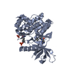
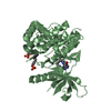
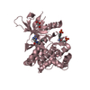
Mass: 419.476 Da / Num. of mol.: 4 / Source method: obtained synthetically / Formula: C23H25N5O3
#3: Chemical ChemComp-GOL / GLYCEROL / GLYCERIN / PROPANE-1,2,3-TRIOL / Glycerol


Mass: 92.094 Da / Num. of mol.: 1 / Source method: obtained synthetically / Formula: C3H8O3
#4: Water ChemComp-HOH / water / Water


Mass: 18.015 Da / Num. of mol.: 279 / Source method: isolated from a natural source / Formula: H2O

-
Experimental details

-
Experiment

ExperimentMethod: X-RAY DIFFRACTION / Number of used crystals: 1

-
Sample preparation

CrystalDensity Matthews: 2.65 Å3/Da / Density % sol: 53.58 % / Description: NONE

-
Data collection

DiffractionMean temperature: 100 K
Diffraction sourceSource: SYNCHROTRON / Site: SLS / Beamline: X06SA / Wavelength: 1
DetectorType: DECTRIS PILATUS 6M / Detector: PIXEL / Date: Feb 16, 2011
RadiationProtocol: SINGLE WAVELENGTH / Monochromatic (M) / Laue (L): M / Scattering type: x-ray
Radiation wavelengthWavelength: 1 Å / Relative weight: 1
ReflectionResolution: 2.35→103.69 Å / Num. obs: 55677 / % possible obs: 97.1 % / Redundancy: 2.9 % / Rmerge(I) obs: 0.1 / Net I/σ(I): 6.9
Reflection shellResolution: 2.35→2.54 Å / Redundancy: 2.7 % / Rmerge(I) obs: 0.45 / Mean I/σ(I) obs: 1.7 / % possible all: 94.3

-
Processing

Software
NameVersionClassification
REFMAC5.6.0117refinement
XDSdata reduction
SCALAdata scaling
REFMACRIGID BODYphasing
RefinementMethod to determine structure: MOLECULAR REPLACEMENT / Resolution: 2.35→103.7 Å / Cor.coef. Fo:Fc: 0.92 / Cor.coef. Fo:Fc free: 0.879 / SU B: 19.145 / SU ML: 0.219 / Cross valid method: THROUGHOUT / ESU R: 0.4 / ESU R Free: 0.263 / Stereochemistry target values: MAXIMUM LIKELIHOOD
Details: HYDROGENS HAVE BEEN ADDED IN THE RIDING POSITIONS. U VALUES WITH TLS ADDED. DISORDERED SIDE CHAINS HAVE OCCUPANCY 0.00
RfactorNum. reflection% reflectionSelection details
Rfree0.26484 924 1.7 %RANDOM
Rwork0.22559 ---
obs0.22627 52795 96.83 %-
Solvent computationIon probe radii: 0.8 Å / Shrinkage radii: 0.8 Å / VDW probe radii: 1.2 Å / Solvent model: BABINET MODEL WITH MASK
Displacement parametersBiso mean: 41.076 Å2
Baniso -1Baniso -2Baniso -3
1--2.55 Å20 Å20.66 Å2
2---2.15 Å20 Å2
3---4.85 Å2
Refinement stepCycle: LAST / Resolution: 2.35→103.7 Å
ProteinNucleic acidLigandSolventTotal
Num. atoms8839 0 130 279 9248
Refine LS restraints
Refine-IDTypeDev idealDev ideal targetNumber
X-RAY DIFFRACTIONr_bond_refined_d0.0080.0199000
X-RAY DIFFRACTIONr_bond_other_d0.0010.026345
X-RAY DIFFRACTIONr_angle_refined_deg1.0982.01312201
X-RAY DIFFRACTIONr_angle_other_deg0.981315267
X-RAY DIFFRACTIONr_dihedral_angle_1_deg5.07851084
X-RAY DIFFRACTIONr_dihedral_angle_2_deg33.63122.853382
X-RAY DIFFRACTIONr_dihedral_angle_3_deg11.645151467
X-RAY DIFFRACTIONr_dihedral_angle_4_deg12.9591575
X-RAY DIFFRACTIONr_chiral_restr0.0570.21318
X-RAY DIFFRACTIONr_gen_planes_refined0.0040.0219885
X-RAY DIFFRACTIONr_gen_planes_other0.0010.021902
X-RAY DIFFRACTIONr_nbd_refined
X-RAY DIFFRACTIONr_nbd_other
X-RAY DIFFRACTIONr_nbtor_refined
X-RAY DIFFRACTIONr_nbtor_other
X-RAY DIFFRACTIONr_xyhbond_nbd_refined
X-RAY DIFFRACTIONr_xyhbond_nbd_other
X-RAY DIFFRACTIONr_metal_ion_refined
X-RAY DIFFRACTIONr_metal_ion_other
X-RAY DIFFRACTIONr_symmetry_vdw_refined
X-RAY DIFFRACTIONr_symmetry_vdw_other
X-RAY DIFFRACTIONr_symmetry_hbond_refined
X-RAY DIFFRACTIONr_symmetry_hbond_other
X-RAY DIFFRACTIONr_symmetry_metal_ion_refined
X-RAY DIFFRACTIONr_symmetry_metal_ion_other
X-RAY DIFFRACTIONr_mcbond_it
X-RAY DIFFRACTIONr_mcbond_other
X-RAY DIFFRACTIONr_mcangle_it
X-RAY DIFFRACTIONr_mcangle_other
X-RAY DIFFRACTIONr_scbond_it
X-RAY DIFFRACTIONr_scbond_other
X-RAY DIFFRACTIONr_scangle_it
X-RAY DIFFRACTIONr_scangle_other
X-RAY DIFFRACTIONr_long_range_B_refined
X-RAY DIFFRACTIONr_long_range_B_other
X-RAY DIFFRACTIONr_rigid_bond_restr
X-RAY DIFFRACTIONr_sphericity_free
X-RAY DIFFRACTIONr_sphericity_bonded
LS refinement shellResolution: 2.353→2.414 Å / Total num. of bins used: 20
RfactorNum. reflection% reflection
Rfree0.382 64 -
Rwork0.29 3527 -
obs--88.97 %
Refinement TLS params.

Method: refined / Refine-ID: X-RAY DIFFRACTION

IDL112)L122)L132)L222)L232)L332)S11 (Å °)S12 (Å °)S13 (Å °)S21 (Å °)S22 (Å °)S23 (Å °)S31 (Å °)S32 (Å °)S33 (Å °)T112)T122)T132)T222)T232)T332)Origin x (Å)Origin y (Å)Origin z (Å)
12.2597-0.3515-0.64325.18791.30752.4571-0.01390.03970.5239-0.33750.1291-0.5781-0.11310.1524-0.1153-0.0842-0.01720.0127-0.04460.0196-0.1062-3.281-8.343-0.117
21.8693-0.1513-0.12122.9489-0.54433.08240.0111-0.1723-0.01210.25460.0498-0.15120.07160.0075-0.0608-0.0936-0.0252-0.0192-0.0449-0.0062-0.1937-15.012-25.29514.428
31.85681.95830.72448.7727-0.13910.9865-0.042-0.0445-0.35780.24990.0218-0.9276-0.02840.20630.0201-0.083-0.0025-0.0439-0.0732-0.0007-0.037819.83727.60456.575
41.4342-0.01140.99123.3973-0.14333.12370.03920.285-0.0109-0.2855-0.0462-0.1859-0.03610.19370.0069-0.09270.02330.0024-0.0476-0.005-0.09896.41640.24338.771
51.27330.9323-0.20226.28370.90031.23170.0301-0.1690.39420.2058-0.10430.86910.0772-0.32410.0742-0.0828-0.00240.0393-0.035-0.0229-0.0252-3.3728.56456.496
61.2912-0.40140.36932.68760.68853.02230.02870.2986-0.0869-0.3328-0.02020.2150.0056-0.0075-0.0084-0.05680.0488-0.0326-0.0339-0.025-0.11449.799-3.55937.865
72.30590.07070.67097.6711.48972.1033-0.0977-0.0036-0.52330.39690.1286-0.85260.06050.2128-0.0309-0.0607-0.0056-0.0322-0.0319-0.005-0.099125.68-47.1640.287
81.97010.11250.5733.30740.20722.4520.03790.20060.1353-0.38340.0376-0.2859-0.10420.1196-0.0754-0.04960.03060.0139-0.02570.0118-0.190613.415-30.323-13.828
Refinement TLS group
IDRefine-IDRefine TLS-IDAuth asym-IDAuth seq-ID
1X-RAY DIFFRACTION1A814 - 904
2X-RAY DIFFRACTION2A905 - 1098
3X-RAY DIFFRACTION3B814 - 904
4X-RAY DIFFRACTION4B905 - 1100
5X-RAY DIFFRACTION5C814 - 904
6X-RAY DIFFRACTION6C905 - 1100
7X-RAY DIFFRACTION7D814 - 904
8X-RAY DIFFRACTION8D905 - 1099

+
About Yorodumi

-
News

-
Feb 9, 2022. New format data for meta-information of EMDB entries

New format data for meta-information of EMDB entries

  • Version 3 of the EMDB header file is now the official format.
  • The previous official version 1.9 will be removed from the archive.

Related info.:EMDB header

External links:wwPDB to switch to version 3 of the EMDB data model

-
Aug 12, 2020. Covid-19 info

Covid-19 info

URL: https://pdbj.org/emnavi/covid19.php

New page: Covid-19 featured information page in EM Navigator.

Related info.:Covid-19 info / Mar 5, 2020. Novel coronavirus structure data

+
Mar 5, 2020. Novel coronavirus structure data

Novel coronavirus structure data

Related info.:Yorodumi Speices / Aug 12, 2020. Covid-19 info

External links:COVID-19 featured content - PDBj / Molecule of the Month (242):Coronavirus Proteases

+
Jan 31, 2019. EMDB accession codes are about to change! (news from PDBe EMDB page)

EMDB accession codes are about to change! (news from PDBe EMDB page)

  • The allocation of 4 digits for EMDB accession codes will soon come to an end. Whilst these codes will remain in use, new EMDB accession codes will include an additional digit and will expand incrementally as the available range of codes is exhausted. The current 4-digit format prefixed with “EMD-” (i.e. EMD-XXXX) will advance to a 5-digit format (i.e. EMD-XXXXX), and so on. It is currently estimated that the 4-digit codes will be depleted around Spring 2019, at which point the 5-digit format will come into force.
  • The EM Navigator/Yorodumi systems omit the EMD- prefix.

Related info.:Q: What is EMD? / ID/Accession-code notation in Yorodumi/EM Navigator

External links:EMDB Accession Codes are Changing Soon! / Contact to PDBj

+
Jul 12, 2017. Major update of PDB

Major update of PDB

  • wwPDB released updated PDB data conforming to the new PDBx/mmCIF dictionary.
  • This is a major update changing the version number from 4 to 5, and with Remediation, in which all the entries are updated.
  • In this update, many items about electron microscopy experimental information are reorganized (e.g. em_software).
  • Now, EM Navigator and Yorodumi are based on the updated data.

External links:wwPDB Remediation / Enriched Model Files Conforming to OneDep Data Standards Now Available in the PDB FTP Archive

-
Yorodumi

Thousand views of thousand structures

  • Yorodumi is a browser for structure data from EMDB, PDB, SASBDB, etc.
  • This page is also the successor to EM Navigator detail page, and also detail information page/front-end page for Omokage search.
  • The word "yorodu" (or yorozu) is an old Japanese word meaning "ten thousand". "mi" (miru) is to see.

Related info.:EMDB / PDB / SASBDB / Comparison of 3 databanks / Yorodumi Search / Aug 31, 2016. New EM Navigator & Yorodumi / Yorodumi Papers / Jmol/JSmol / Function and homology information / Changes in new EM Navigator and Yorodumi

Read more