[English] 日本語
Yorodumi
- PDB-5mev: MCL1 FAB COMPLEX IN COMPLEX WITH COMPOUND 21 -

+
Open data


ID or keywords:

Loading...

-
Basic information

Entry
Database: PDB / ID: 5mev
TitleMCL1 FAB COMPLEX IN COMPLEX WITH COMPOUND 21
Components
  • Fab Heavy ChainFragment antigen-binding
  • Fab Light ChainFragment antigen-binding
  • Induced myeloid leukemia cell differentiation protein Mcl-1 homolog,Induced myeloid leukemia cell differentiation protein Mcl-1
KeywordsIMMUNE SYSTEM / MCL1 FAB macrocycle / MCL1-FAB_55_C6HIS
Function / homology
Function and homology information


BH domain binding / positive regulation of oxidative stress-induced neuron intrinsic apoptotic signaling pathway / cellular homeostasis / cell fate determination / mitochondrial fusion / channel activity / Bcl-2 family protein complex / BH3 domain binding / negative regulation of anoikis / protein transmembrane transporter activity ...BH domain binding / positive regulation of oxidative stress-induced neuron intrinsic apoptotic signaling pathway / cellular homeostasis / cell fate determination / mitochondrial fusion / channel activity / Bcl-2 family protein complex / BH3 domain binding / negative regulation of anoikis / protein transmembrane transporter activity / negative regulation of extrinsic apoptotic signaling pathway in absence of ligand / extrinsic apoptotic signaling pathway in absence of ligand / negative regulation of autophagy / release of cytochrome c from mitochondria / response to cytokine / Signaling by ALK fusions and activated point mutants / intrinsic apoptotic signaling pathway in response to DNA damage / regulation of apoptotic process / Interleukin-4 and Interleukin-13 signaling / mitochondrial outer membrane / cell differentiation / protein dimerization activity / positive regulation of apoptotic process / protein heterodimerization activity / DNA damage response / protein-containing complex binding / negative regulation of apoptotic process / protein homodimerization activity / mitochondrion / nucleoplasm / membrane / nucleus / cytosol / cytoplasm
Similarity search - Function
Apoptosis regulator, Mcl-1 / Blc2-like / Apoptosis Regulator Bcl-x / Apoptosis regulator, Bcl-2, BH3 motif, conserved site / Apoptosis regulator, Bcl-2 family BH3 motif signature. / Apoptosis regulator, Bcl-2, BH1 motif, conserved site / Apoptosis regulator, Bcl-2 family BH1 motif signature. / Apoptosis regulator, Bcl-2, BH2 motif, conserved site / Apoptosis regulator, Bcl-2 family BH2 motif signature. / BCL (B-Cell lymphoma); contains BH1, BH2 regions ...Apoptosis regulator, Mcl-1 / Blc2-like / Apoptosis Regulator Bcl-x / Apoptosis regulator, Bcl-2, BH3 motif, conserved site / Apoptosis regulator, Bcl-2 family BH3 motif signature. / Apoptosis regulator, Bcl-2, BH1 motif, conserved site / Apoptosis regulator, Bcl-2 family BH1 motif signature. / Apoptosis regulator, Bcl-2, BH2 motif, conserved site / Apoptosis regulator, Bcl-2 family BH2 motif signature. / BCL (B-Cell lymphoma); contains BH1, BH2 regions / Bcl-2 family / Bcl-2, Bcl-2 homology region 1-3 / Bcl2-like / Apoptosis regulator proteins, Bcl-2 family / BCL2-like apoptosis inhibitors family profile. / Bcl-2-like superfamily / Immunoglobulins / Immunoglobulin-like / Sandwich / Orthogonal Bundle / Mainly Beta / Mainly Alpha
Similarity search - Domain/homology
Chem-7LW / Induced myeloid leukemia cell differentiation protein Mcl-1 homolog / Induced myeloid leukemia cell differentiation protein Mcl-1
Similarity search - Component
Biological speciesMus musculus (house mouse)
Homo sapiens (human)
MethodX-RAY DIFFRACTION / SYNCHROTRON / Resolution: 2.94 Å
AuthorsHargreaves, D.
CitationJournal: Acs Med.Chem.Lett. / Year: 2017
Title: Structure Based Design of Non-Natural Peptidic Macrocyclic Mcl-1 Inhibitors
Authors: Johannes, J.W. / Bates, S. / Beigie, C. / Belmonte, M. / Breen, J. / Cao, S. / Centrella, P.A. / Clark, M.A. / Cuozzo, J.W. / Dumelin, C.E. / Ferguson, A.D. / Habeshian, S. / Hargreaves, D. ...Authors: Johannes, J.W. / Bates, S. / Beigie, C. / Belmonte, M. / Breen, J. / Cao, S. / Centrella, P.A. / Clark, M.A. / Cuozzo, J.W. / Dumelin, C.E. / Ferguson, A.D. / Habeshian, S. / Hargreaves, D. / Joubran, C. / Kazmirski, S. / Keefe, A. / Lamb, M.L. / Lan, H. / Li, Y. / Ma, H. / Mlynarski, S. / Packer, M.J. / Rawlins, P. / Robbins, D.W. / Shen, H. / Sigel, E.A. / Soutter, H.H. / Su, N. / Trost, D.M. / Wang, H. / Wickson, K.F. / Wu, C. / Zhang, Y. / Zhao, Q. / Zheng, X. / Hird, A.
History
DepositionNov 16, 2016Deposition site: PDBE / Processing site: PDBE
Revision 1.0Jan 18, 2017Provider: repository / Type: Initial release
Revision 1.1Feb 22, 2017Group: Database references
Revision 1.2Mar 8, 2017Group: Database references

-
Structure visualization

Structure viewerMolecule:
MolmilJmol/JSmol

Downloads & links

-
Assembly

Deposited unit
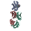
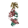
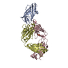
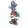
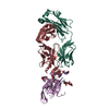
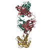
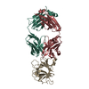
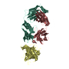
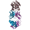
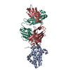
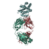
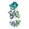
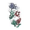
A: Induced myeloid leukemia cell differentiation protein Mcl-1 homolog,Induced myeloid leukemia cell differentiation protein Mcl-1
H: Fab Heavy Chain
L: Fab Light Chain
hetero molecules


Theoretical massNumber of molelcules
Total (without water)67,6304
Polymers66,8743
Non-polymers7571
Water1,49583
1


  • Idetical with deposited unit
  • defined by author&software
TypeNameSymmetry operationNumber
identity operation1_555x,y,z1
Buried area5000 Å2
ΔGint-28 kcal/mol
Surface area24900 Å2
MethodPISA
Unit cell
Length a, b, c (Å)166.514, 39.705, 103.515
Angle α, β, γ (deg.)90.00, 125.74, 90.00
Int Tables number5
Space group name H-MC121

-
Components

#1: Protein Induced myeloid leukemia cell differentiation protein Mcl-1 homolog,Induced myeloid leukemia cell differentiation protein Mcl-1 / Bcl-2-related protein EAT/mcl1 / Bcl-2-like protein 3 / Bcl2-L-3 / Bcl-2-related protein EAT/mcl1 / mcl1/EAT


Mass: 18227.592 Da / Num. of mol.: 1
Source method: isolated from a genetically manipulated source
Source: (gene. exp.) Mus musculus (house mouse), (gene. exp.) Homo sapiens (human)
Gene: Mcl1, MCL1, BCL2L3 / Production host: Escherichia coli (E. coli) / References: UniProt: P97287, UniProt: Q07820
#2: Antibody Fab Heavy Chain / Fragment antigen-binding


Mass: 24488.490 Da / Num. of mol.: 1
Source method: isolated from a genetically manipulated source
Source: (gene. exp.) Homo sapiens (human) / Production host: Escherichia coli (E. coli)
#3: Antibody Fab Light Chain / Fragment antigen-binding


Mass: 24157.469 Da / Num. of mol.: 1
Source method: isolated from a genetically manipulated source
Source: (gene. exp.) Homo sapiens (human) / Production host: Escherichia coli (E. coli)
#4: Chemical ChemComp-7LW / (5~{R},13~{S},17~{S})-5-[(3,4-dichlorophenyl)methyl]-8-methyl-13-[(4-methylsulfonylphenyl)methyl]-1,4,8,12,16-pentazatricyclo[15.8.1.0^{20,25}]hexacosa-20,22,24-triene-3,7,15,26-tetrone


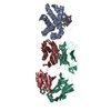
Mass: 756.738 Da / Num. of mol.: 1 / Source method: obtained synthetically / Formula: C37H43Cl2N5O6S
#5: Water ChemComp-HOH / water / Water


Mass: 18.015 Da / Num. of mol.: 83 / Source method: isolated from a natural source / Formula: H2O

-
Experimental details

-
Experiment

ExperimentMethod: X-RAY DIFFRACTION / Number of used crystals: 1

-
Sample preparation

CrystalDensity Matthews: 2.06 Å3/Da / Density % sol: 40.41 %
Crystal growTemperature: 293 K / Method: vapor diffusion, sitting drop / Details: PEG 8K 10%w/v, PEG 1500 10%w/v

-
Data collection

DiffractionMean temperature: 100 K
Diffraction sourceSource: SYNCHROTRON / Site: Diamond / Beamline: I03 / Wavelength: 0.97626 Å
DetectorType: DECTRIS PILATUS3 X 1M / Detector: PIXEL / Date: May 16, 2015
RadiationProtocol: SINGLE WAVELENGTH / Monochromatic (M) / Laue (L): M / Scattering type: x-ray
Radiation wavelength
IDWavelength (Å)Relative weight
10.976251
20.976261
ReflectionResolution: 2.94→28.9 Å / Num. obs: 11919 / % possible obs: 98.4 % / Redundancy: 3.2 % / Biso Wilson estimate: 83.87 Å2 / CC1/2: 0.974 / Rmerge(I) obs: 0.134 / Rpim(I) all: 0.128 / Rrim(I) all: 0.195 / Net I/σ(I): 6.2
Reflection shellResolution: 2.94→3.01 Å / Redundancy: 3.1 % / Rmerge(I) obs: 0.637 / Mean I/σ(I) obs: 1.1 / CC1/2: 0.375 / % possible all: 89

-
Processing

Software
NameVersionClassification
REFMAC5.8.0135refinement
MOSFLMdata reduction
SCALAdata scaling
PHASERphasing
RefinementResolution: 2.94→26.45 Å / Cor.coef. Fo:Fc: 0.921 / Cor.coef. Fo:Fc free: 0.864 / SU B: 23.329 / SU ML: 0.424 / Cross valid method: THROUGHOUT / ESU R Free: 0.572 / Stereochemistry target values: MAXIMUM LIKELIHOOD / Details: HYDROGENS HAVE BEEN ADDED IN THE RIDING POSITIONS
RfactorNum. reflection% reflectionSelection details
Rfree0.30248 597 5 %RANDOM
Rwork0.22502 ---
obs0.229 11314 98.63 %-
Solvent computationIon probe radii: 0.8 Å / Shrinkage radii: 0.8 Å / VDW probe radii: 1.2 Å / Solvent model: MASK
Displacement parametersBiso mean: 56.54 Å2
Baniso -1Baniso -2Baniso -3
1-0.65 Å2-0 Å20.21 Å2
2---0.6 Å2-0 Å2
3----0.18 Å2
Refinement stepCycle: 1 / Resolution: 2.94→26.45 Å
ProteinNucleic acidLigandSolventTotal
Num. atoms4354 0 51 83 4488
Refine LS restraints
Refine-IDTypeDev idealDev ideal targetNumber
X-RAY DIFFRACTIONr_bond_refined_d0.0080.0194505
X-RAY DIFFRACTIONr_bond_other_d0.0020.024184
X-RAY DIFFRACTIONr_angle_refined_deg1.3291.946125
X-RAY DIFFRACTIONr_angle_other_deg0.92839653
X-RAY DIFFRACTIONr_dihedral_angle_1_deg6.6055566
X-RAY DIFFRACTIONr_dihedral_angle_2_deg39.74423.729177
X-RAY DIFFRACTIONr_dihedral_angle_3_deg18.32715720
X-RAY DIFFRACTIONr_dihedral_angle_4_deg15.2661526
X-RAY DIFFRACTIONr_chiral_restr0.0680.2691
X-RAY DIFFRACTIONr_gen_planes_refined0.0040.0215078
X-RAY DIFFRACTIONr_gen_planes_other0.0010.021018
X-RAY DIFFRACTIONr_nbd_refined
X-RAY DIFFRACTIONr_nbd_other
X-RAY DIFFRACTIONr_nbtor_refined
X-RAY DIFFRACTIONr_nbtor_other
X-RAY DIFFRACTIONr_xyhbond_nbd_refined
X-RAY DIFFRACTIONr_xyhbond_nbd_other
X-RAY DIFFRACTIONr_metal_ion_refined
X-RAY DIFFRACTIONr_metal_ion_other
X-RAY DIFFRACTIONr_symmetry_vdw_refined
X-RAY DIFFRACTIONr_symmetry_vdw_other
X-RAY DIFFRACTIONr_symmetry_hbond_refined
X-RAY DIFFRACTIONr_symmetry_hbond_other
X-RAY DIFFRACTIONr_symmetry_metal_ion_refined
X-RAY DIFFRACTIONr_symmetry_metal_ion_other
X-RAY DIFFRACTIONr_mcbond_it2.0375.6472280
X-RAY DIFFRACTIONr_mcbond_other2.0345.6462278
X-RAY DIFFRACTIONr_mcangle_it3.4578.4612840
X-RAY DIFFRACTIONr_mcangle_other3.4568.4622841
X-RAY DIFFRACTIONr_scbond_it1.9635.752225
X-RAY DIFFRACTIONr_scbond_other1.9625.7512226
X-RAY DIFFRACTIONr_scangle_it
X-RAY DIFFRACTIONr_scangle_other3.4028.533286
X-RAY DIFFRACTIONr_long_range_B_refined6.56243.2514897
X-RAY DIFFRACTIONr_long_range_B_other6.54543.2814889
X-RAY DIFFRACTIONr_rigid_bond_restr
X-RAY DIFFRACTIONr_sphericity_free
X-RAY DIFFRACTIONr_sphericity_bonded
LS refinement shellResolution: 2.94→3.016 Å / Total num. of bins used: 20
RfactorNum. reflection% reflection
Rfree0.552 35 -
Rwork0.415 784 -
obs--94.35 %

+
About Yorodumi

-
News

-
Feb 9, 2022. New format data for meta-information of EMDB entries

New format data for meta-information of EMDB entries

  • Version 3 of the EMDB header file is now the official format.
  • The previous official version 1.9 will be removed from the archive.

Related info.:EMDB header

External links:wwPDB to switch to version 3 of the EMDB data model

-
Aug 12, 2020. Covid-19 info

Covid-19 info

URL: https://pdbj.org/emnavi/covid19.php

New page: Covid-19 featured information page in EM Navigator.

Related info.:Covid-19 info / Mar 5, 2020. Novel coronavirus structure data

+
Mar 5, 2020. Novel coronavirus structure data

Novel coronavirus structure data

Related info.:Yorodumi Speices / Aug 12, 2020. Covid-19 info

External links:COVID-19 featured content - PDBj / Molecule of the Month (242):Coronavirus Proteases

+
Jan 31, 2019. EMDB accession codes are about to change! (news from PDBe EMDB page)

EMDB accession codes are about to change! (news from PDBe EMDB page)

  • The allocation of 4 digits for EMDB accession codes will soon come to an end. Whilst these codes will remain in use, new EMDB accession codes will include an additional digit and will expand incrementally as the available range of codes is exhausted. The current 4-digit format prefixed with “EMD-” (i.e. EMD-XXXX) will advance to a 5-digit format (i.e. EMD-XXXXX), and so on. It is currently estimated that the 4-digit codes will be depleted around Spring 2019, at which point the 5-digit format will come into force.
  • The EM Navigator/Yorodumi systems omit the EMD- prefix.

Related info.:Q: What is EMD? / ID/Accession-code notation in Yorodumi/EM Navigator

External links:EMDB Accession Codes are Changing Soon! / Contact to PDBj

+
Jul 12, 2017. Major update of PDB

Major update of PDB

  • wwPDB released updated PDB data conforming to the new PDBx/mmCIF dictionary.
  • This is a major update changing the version number from 4 to 5, and with Remediation, in which all the entries are updated.
  • In this update, many items about electron microscopy experimental information are reorganized (e.g. em_software).
  • Now, EM Navigator and Yorodumi are based on the updated data.

External links:wwPDB Remediation / Enriched Model Files Conforming to OneDep Data Standards Now Available in the PDB FTP Archive

-
Yorodumi

Thousand views of thousand structures

  • Yorodumi is a browser for structure data from EMDB, PDB, SASBDB, etc.
  • This page is also the successor to EM Navigator detail page, and also detail information page/front-end page for Omokage search.
  • The word "yorodu" (or yorozu) is an old Japanese word meaning "ten thousand". "mi" (miru) is to see.

Related info.:EMDB / PDB / SASBDB / Comparison of 3 databanks / Yorodumi Search / Aug 31, 2016. New EM Navigator & Yorodumi / Yorodumi Papers / Jmol/JSmol / Function and homology information / Changes in new EM Navigator and Yorodumi

Read more