 Movie
Movie Controller


+ Open data

- Basic information
| Entry | Database: PDB / ID: 6qb6 | ||||||
|---|---|---|---|---|---|---|---|
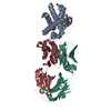
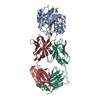
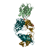
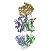
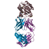
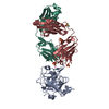
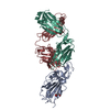
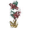
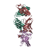
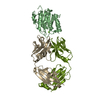
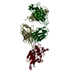
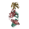
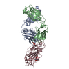
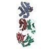
| Title | Mcl1 in complex with a Fab | ||||||
 Components Components |
| ||||||
 Keywords Keywords |  APOPTOSIS / Mcl1 / Fab / AZD5991 APOPTOSIS / Mcl1 / Fab / AZD5991 | ||||||
| Function / homology |  Function and homology information Function and homology informationpositive regulation of oxidative stress-induced neuron intrinsic apoptotic signaling pathway / cellular homeostasis / cell fate determination / channel activity / mitochondrial fusion / Bcl-2 family protein complex / BH3 domain binding / protein transmembrane transporter activity / negative regulation of anoikis / negative regulation of extrinsic apoptotic signaling pathway in absence of ligand ...positive regulation of oxidative stress-induced neuron intrinsic apoptotic signaling pathway / cellular homeostasis / cell fate determination / channel activity / mitochondrial fusion / Bcl-2 family protein complex / BH3 domain binding / protein transmembrane transporter activity / negative regulation of anoikis / negative regulation of extrinsic apoptotic signaling pathway in absence of ligand / extrinsic apoptotic signaling pathway in absence of ligand / negative regulation of autophagy / release of cytochrome c from mitochondria / response to cytokine / intrinsic apoptotic signaling pathway in response to DNA damage / Interleukin-4 and Interleukin-13 signaling / regulation of apoptotic process / mitochondrial outer membrane / positive regulation of apoptotic process / protein heterodimerization activity / DNA damage response / negative regulation of apoptotic process / protein homodimerization activity / mitochondrion / nucleoplasm / membrane / nucleus / cytosol / cytoplasmSimilarity search - Function | ||||||
| Biological species |  Homo sapiens (human) Homo sapiens (human) | ||||||
| Method | X-RAY DIFFRACTION / SYNCHROTRON / MOLECULAR REPLACEMENT / Resolution: 2.24 Å | ||||||
 Authors Authors | Hargreaves, D. | ||||||
 Citation Citation | Journal: Acta Crystallogr D Struct Biol / Year: 2019 Title: Antibody fragments structurally enable a drug-discovery campaign on the cancer target Mcl-1. Authors: Luptak, J. / Bista, M. / Fisher, D. / Flavell, L. / Gao, N. / Wickson, K. / Kazmirski, S.L. / Howard, T. / Rawlins, P.B. / Hargreaves, D. | ||||||
| History |
|
- Structure visualization
| Structure viewer | Molecule: MolmilJmol/JSmol |
|---|
- Downloads & links
-Download
| PDBx/mmCIF format | 6qb6.cif.gz | 125.3 KB | Display | PDBx/mmCIF format |
|---|---|---|---|---|
| PDB format | pdb6qb6.ent.gz | 99.2 KB | Display | PDB format |
| PDBx/mmJSON format | 6qb6.json.gz | Tree view | PDBx/mmJSON format | |
| Others |  Other downloads Other downloads |
-Validation report
| Arichive directory | https://data.pdbj.org/pub/pdb/validation_reports/qb/6qb6ftp://data.pdbj.org/pub/pdb/validation_reports/qb/6qb6 | HTTPS FTP |
|---|
-Related structure data
| Related structure data |  6qb3C  6qb4C  6qb9C  6qbcC  6qf9C 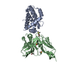 6qfcC C: citing same article ( |
|---|---|
| Similar structure data |


-Links
 PDBj
PDBj


- Assembly
| Deposited unit | 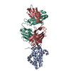
| ||||||||
|---|---|---|---|---|---|---|---|---|---|
| 1 |
| ||||||||
| Unit cell |
|
-Components
| #1: Protein | Mass: 18227.592 Da / Num. of mol.: 1 Source method: isolated from a genetically manipulated source Source: (gene. exp.) Homo sapiens (human) / Gene: MCL1, BCL2L3 / Production host:  Escherichia coli (E. coli) / References: UniProt: Q07820 Escherichia coli (E. coli) / References: UniProt: Q07820 |
|---|---|
| #2: Antibody | Fragment antigen-binding Mass: 24488.490 Da / Num. of mol.: 1 Source method: isolated from a genetically manipulated source Source: (gene. exp.) Homo sapiens (human) / Production host:   Cricetulus griseus (Chinese hamster) Cricetulus griseus (Chinese hamster) |
| #3: Antibody | Fragment antigen-binding Mass: 23494.688 Da / Num. of mol.: 1 Source method: isolated from a genetically manipulated source Source: (gene. exp.) Homo sapiens (human) / Production host: Cricetulus griseus (Chinese hamster) |
| #4: Water | ChemComp-HOH / Water Mass: 18.015 Da / Num. of mol.: 154 / Source method: isolated from a natural source / Formula: H2O Mass: 18.015 Da / Num. of mol.: 154 / Source method: isolated from a natural source / Formula: H2O |
-Experimental details
-Experiment
| Experiment | Method: X-RAY DIFFRACTION / Number of used crystals: 1 |
|---|
- Sample preparation
| Crystal | Density Matthews: 2.32 Å3/Da / Density % sol: 46.93 % |
|---|---|
| Crystal grow | Temperature: 293 K / Method: vapor diffusion, sitting drop / Details: 54.5 mM PCTP4 45.5 mM PCTP10 15 %w/v PEG-2000 MME |
-Data collection
| Diffraction | Mean temperature: 100 K / Serial crystal experiment: N |
|---|---|
| Diffraction source | Source: SYNCHROTRON / Site: Diamond  / Beamline: I02 / Wavelength: 0.979 Å / Beamline: I02 / Wavelength: 0.979 Å |
| Detector | Type: ADSC QUANTUM 315 / Detector: CCD / Date: Jul 11, 2013 |
| Radiation | Protocol: SINGLE WAVELENGTH / Monochromatic (M) / Laue (L): M / Scattering type: x-ray |
| Radiation wavelength | Wavelength: 0.979 Å / Relative weight: 1 |
| Reflection | Resolution: 2.24→50.1 Å / Num. obs: 29461 / % possible obs: 99.3 % / Redundancy: 3.3 % / Biso Wilson estimate: 43.92 Å2 / Net I/σ(I): 12.3 |
| Reflection shell | Resolution: 2.24→2.3 Å |
- Processing
| Software |
| ||||||||||||||||||||||||||||||||||||||||||||||||||||||||||||||||||||||||||||||||||||||||||||||||||||||||||||||||||
|---|---|---|---|---|---|---|---|---|---|---|---|---|---|---|---|---|---|---|---|---|---|---|---|---|---|---|---|---|---|---|---|---|---|---|---|---|---|---|---|---|---|---|---|---|---|---|---|---|---|---|---|---|---|---|---|---|---|---|---|---|---|---|---|---|---|---|---|---|---|---|---|---|---|---|---|---|---|---|---|---|---|---|---|---|---|---|---|---|---|---|---|---|---|---|---|---|---|---|---|---|---|---|---|---|---|---|---|---|---|---|---|---|---|---|---|
| Refinement | Method to determine structure: MOLECULAR REPLACEMENT / Resolution: 2.24→47.71 Å / Cor.coef. Fo:Fc: 0.942 / Cor.coef. Fo:Fc free: 0.911 / SU R Cruickshank DPI: 0.269 / Cross valid method: THROUGHOUT / σ(F): 0 / SU R Blow DPI: 0.268 / SU Rfree Blow DPI: 0.202 / SU Rfree Cruickshank DPI: 0.205
| ||||||||||||||||||||||||||||||||||||||||||||||||||||||||||||||||||||||||||||||||||||||||||||||||||||||||||||||||||
| Displacement parameters | Biso mean: 47.2 Å2
| ||||||||||||||||||||||||||||||||||||||||||||||||||||||||||||||||||||||||||||||||||||||||||||||||||||||||||||||||||
| Refine analyze | Luzzati coordinate error obs: 0.28 Å | ||||||||||||||||||||||||||||||||||||||||||||||||||||||||||||||||||||||||||||||||||||||||||||||||||||||||||||||||||
| Refinement step | Cycle: 1 / Resolution: 2.24→47.71 Å
| ||||||||||||||||||||||||||||||||||||||||||||||||||||||||||||||||||||||||||||||||||||||||||||||||||||||||||||||||||
| Refine LS restraints |
| ||||||||||||||||||||||||||||||||||||||||||||||||||||||||||||||||||||||||||||||||||||||||||||||||||||||||||||||||||
| LS refinement shell | Resolution: 2.24→2.26 Å / Total num. of bins used: 50
|
