 Movie
Movie Controller

[English] 日本語
 Yorodumi
- PDB-6bkc: Structure of Hepatitis C Virus Envelope Glycoprotein E2 core from... -
+ Open data

- Basic information
| Entry | Database: PDB / ID: 6bkc | |||||||||
|---|---|---|---|---|---|---|---|---|---|---|
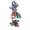
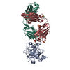
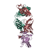
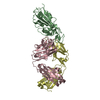
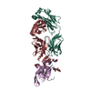
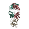
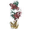
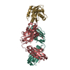
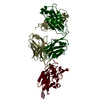
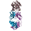
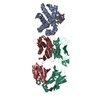
| Title | Structure of Hepatitis C Virus Envelope Glycoprotein E2 core from genotype 6a bound to broadly neutralizing antibody AR3B | |||||||||
 Components Components |
| |||||||||
 Keywords Keywords |  IMMUNE SYSTEM / HCV / broadly neutralizing antibodies / bNAbs / E2 core / IGHV1-69 IMMUNE SYSTEM / HCV / broadly neutralizing antibodies / bNAbs / E2 core / IGHV1-69 | |||||||||
| Function / homology |  Function and homology information Function and homology informationhost cell mitochondrial membrane / host cell lipid droplet / symbiont-mediated suppression of host TRAF-mediated signal transduction / transformation of host cell by virus / symbiont-mediated perturbation of host cell cycle G1/S transition checkpoint / immunoglobulin complex, circulating / immunoglobulin receptor binding / symbiont-mediated suppression of host JAK-STAT cascade via inhibition of STAT1 activity / : / symbiont-mediated suppression of host cytoplasmic pattern recognition receptor signaling pathway via inhibition of MAVS activity ...host cell mitochondrial membrane / host cell lipid droplet / symbiont-mediated suppression of host TRAF-mediated signal transduction / transformation of host cell by virus / symbiont-mediated perturbation of host cell cycle G1/S transition checkpoint / immunoglobulin complex, circulating / immunoglobulin receptor binding / symbiont-mediated suppression of host JAK-STAT cascade via inhibition of STAT1 activity / : / symbiont-mediated suppression of host cytoplasmic pattern recognition receptor signaling pathway via inhibition of MAVS activity / ribonucleoside triphosphate phosphatase activity / lipid droplet / complement activation, classical pathway / antigen binding / protein complex oligomerization / monoatomic ion channel activity / antibacterial humoral response / viral nucleocapsid / clathrin-dependent endocytosis of virus by host cell / blood microparticle / RNA helicase activity / host cell perinuclear region of cytoplasm / host cell endoplasmic reticulum membrane / symbiont-mediated suppression of host type I interferon-mediated signaling pathway / ribonucleoprotein complex / induction by virus of host autophagy / immune response / viral RNA genome replication / cysteine-type endopeptidase activity / RNA-dependent RNA polymerase activity / serine-type endopeptidase activity / fusion of virus membrane with host endosome membrane / viral envelope / host cell nucleus / virion attachment to host cell / host cell plasma membrane / virion membrane / structural molecule activity / proteolysis / extracellular space / RNA binding / extracellular exosome / zinc ion binding / ATP binding / metal ion binding / plasma membrane / cytoplasmSimilarity search - Function | |||||||||
| Biological species |  Homo sapiens (human) Homo sapiens (human) Recombinant Hepatitis C virus HK6a/JFH-1 Recombinant Hepatitis C virus HK6a/JFH-1 | |||||||||
| Method | X-RAY DIFFRACTION / SYNCHROTRON / MOLECULAR REPLACEMENT / Resolution: 2.6 Å | |||||||||
 Authors Authors | Tzarum, N. / Wilson, I.A. / Law, M. | |||||||||
| Funding support |  United States, 2items United States, 2items
| |||||||||
 Citation Citation | Journal: Sci Adv / Year: 2019 Title: Genetic and structural insights into broad neutralization of hepatitis C virus by human VH1-69 antibodies. Authors: Tzarum, N. / Giang, E. / Kong, L. / He, L. / Prentoe, J. / Augestad, E. / Hua, Y. / Castillo, S. / Lauer, G.M. / Bukh, J. / Zhu, J. / Wilson, I.A. / Law, M. | |||||||||
| History |
|
- Structure visualization
| Structure viewer | Molecule: MolmilJmol/JSmol |
|---|
- Downloads & links
-Download
| PDBx/mmCIF format | 6bkc.cif.gz | 128.4 KB | Display | PDBx/mmCIF format |
|---|---|---|---|---|
| PDB format | pdb6bkc.ent.gz | 95.4 KB | Display | PDB format |
| PDBx/mmJSON format | 6bkc.json.gz | Tree view | PDBx/mmJSON format | |
| Others |  Other downloads Other downloads |
-Validation report
| Arichive directory | https://data.pdbj.org/pub/pdb/validation_reports/bk/6bkcftp://data.pdbj.org/pub/pdb/validation_reports/bk/6bkc | HTTPS FTP |
|---|
-Related structure data
| Related structure data |  6bkbC 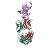 6bkdC  6kbk S: Starting model for refinement C: citing same article ( |
|---|---|
| Similar structure data |
-Links
 PDBj
PDBj


- Assembly
| Deposited unit | 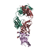
| ||||||||
|---|---|---|---|---|---|---|---|---|---|
| 1 |
| ||||||||
| Unit cell |
|
-Components
| #1: Antibody | Mass: 24035.156 Da / Num. of mol.: 1 Source method: isolated from a genetically manipulated source Source: (gene. exp.) Homo sapiens (human) / Production host: Homo sapiens (human) / References: UniProt: P0DOX5 | ||
|---|---|---|---|
| #2: Antibody | Mass: 23418.855 Da / Num. of mol.: 1 Source method: isolated from a genetically manipulated source Source: (gene. exp.) Homo sapiens (human) / Production host: Homo sapiens (human) / References: UniProt: Q8TCD0 | ||
| #3: Protein | Proteolysis Mass: 20707.395 Da / Num. of mol.: 1 / Mutation: N448D Source method: isolated from a genetically manipulated source Source: (gene. exp.) Recombinant Hepatitis C virus HK6a/JFH-1Production host: Homo sapiens (human) / References: UniProt: B9V0E2 | ||
| #4: Sugar | ChemComp-NAG / N-Acetylglucosamine  Type: D-saccharide, beta linking / Mass: 221.208 Da / Num. of mol.: 4 Type: D-saccharide, beta linking / Mass: 221.208 Da / Num. of mol.: 4Source method: isolated from a genetically manipulated source Formula: C8H15NO6 #5: Water | ChemComp-HOH / | Water Mass: 18.015 Da / Num. of mol.: 47 / Source method: isolated from a natural source / Formula: H2O Mass: 18.015 Da / Num. of mol.: 47 / Source method: isolated from a natural source / Formula: H2O |
-Experimental details
-Experiment
| Experiment | Method: X-RAY DIFFRACTION / Number of used crystals: 1 |
|---|
- Sample preparation
| Crystal | Density Matthews: 2.29 Å3/Da / Density % sol: 46.22 % |
|---|---|
| Crystal grow | Temperature: 293 K / Method: vapor diffusion, sitting drop / pH: 6.7 / Details: 20% (w/v) PEG 3500, 0.2M Li-chloride |
-Data collection
| Diffraction | Mean temperature: 100 K / Serial crystal experiment: N |
|---|---|
| Diffraction source | Source: SYNCHROTRON / Site: SSRL / Beamline: BL12-2 / Wavelength: 0.97946 Å |
| Detector | Type: DECTRIS PILATUS3 S 6M / Detector: PIXEL / Date: Jul 22, 2016 |
| Radiation | Protocol: SINGLE WAVELENGTH / Monochromatic (M) / Laue (L): M / Scattering type: x-ray |
| Radiation wavelength | Wavelength: 0.97946 Å / Relative weight: 1 |
| Reflection | Resolution: 2.434→35 Å / Num. obs: 20780 / % possible obs: 90.1 % / Redundancy: 3.1 % / Rpim(I) all: 0.06 / Rsym value: 0.12 / Net I/σ(I): 10.2 |
| Reflection shell | Resolution: 2.45→2.49 Å / Redundancy: 1.8 % / Mean I/σ(I) obs: 1.6 / Num. unique obs: 815 / CC1/2: 0.66 / % possible all: 59.2 |
- Processing
| Software |
| ||||||||||||||||||||||||||||||||||||||||||||||||||||||||
|---|---|---|---|---|---|---|---|---|---|---|---|---|---|---|---|---|---|---|---|---|---|---|---|---|---|---|---|---|---|---|---|---|---|---|---|---|---|---|---|---|---|---|---|---|---|---|---|---|---|---|---|---|---|---|---|---|---|
| Refinement | Method to determine structure: MOLECULAR REPLACEMENT Starting model: 6kbk 6kbk Resolution: 2.6→32.58 Å / SU ML: 0.37 / Cross valid method: FREE R-VALUE / σ(F): 1.35 / Phase error: 29.45 / Stereochemistry target values: ML
| ||||||||||||||||||||||||||||||||||||||||||||||||||||||||
| Solvent computation | Shrinkage radii: 0.9 Å / VDW probe radii: 1.11 Å / Solvent model: FLAT BULK SOLVENT MODEL | ||||||||||||||||||||||||||||||||||||||||||||||||||||||||
| Refinement step | Cycle: LAST / Resolution: 2.6→32.58 Å
| ||||||||||||||||||||||||||||||||||||||||||||||||||||||||
| Refine LS restraints |
| ||||||||||||||||||||||||||||||||||||||||||||||||||||||||
| LS refinement shell |
|
