 Movie
Movie Controller


+ Open data

- Basic information
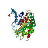
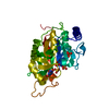
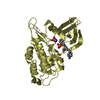
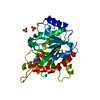
| Entry | Database: PDB / ID: 5lru | |||||||||
|---|---|---|---|---|---|---|---|---|---|---|
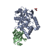
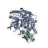
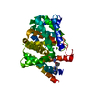
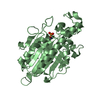
| Title | Structure of Cezanne/OTUD7B OTU domain | |||||||||
 Components Components | OTU domain-containing protein 7B | |||||||||
 Keywords Keywords |  HYDROLASE / protease / deubiquitinase / OTU domain HYDROLASE / protease / deubiquitinase / OTU domain | |||||||||
| Function / homology |  Function and homology information Function and homology informationmucosal immune response / protein K11-linked deubiquitination / negative regulation of interleukin-8 production / protein K48-linked deubiquitination / protein K63-linked deubiquitination / negative regulation of protein localization to nucleus / K48-linked deubiquitinase activity / TNFR1-induced proapoptotic signaling / K63-linked polyubiquitin modification-dependent protein binding / negative regulation of canonical NF-kappaB signal transduction ...mucosal immune response / protein K11-linked deubiquitination / negative regulation of interleukin-8 production / protein K48-linked deubiquitination / protein K63-linked deubiquitination / negative regulation of protein localization to nucleus / K48-linked deubiquitinase activity / TNFR1-induced proapoptotic signaling / K63-linked polyubiquitin modification-dependent protein binding / negative regulation of canonical NF-kappaB signal transduction / cysteine-type peptidase activity / TNFR1-induced NF-kappa-B signaling pathway / Regulation of TNFR1 signaling / Ovarian tumor domain proteases / in utero embryonic development / adaptive immune response / ubiquitinyl hydrolase 1 / cysteine-type deubiquitinase activity / negative regulation of transcription by RNA polymerase II / proteolysis / DNA binding / zinc ion binding / nucleus / cytosol / cytoplasmSimilarity search - Function | |||||||||
| Biological species |  Homo sapiens (human) Homo sapiens (human) | |||||||||
| Method | X-RAY DIFFRACTION / SYNCHROTRON / MOLECULAR REPLACEMENT / Resolution: 2.2 Å | |||||||||
 Authors Authors | Mevissen, T.E.T. / Kulathu, Y. / Mulder, M.P.C. / Geurink, P.P. / Maslen, S.L. / Gersch, M. / Elliott, P.R. / Burke, J.E. / van Tol, B.D.M. / Akutsu, M. ...Mevissen, T.E.T. / Kulathu, Y. / Mulder, M.P.C. / Geurink, P.P. / Maslen, S.L. / Gersch, M. / Elliott, P.R. / Burke, J.E. / van Tol, B.D.M. / Akutsu, M. / El Oualid, F. / Kawasaki, M. / Freund, S.M.V. / Ovaa, H. / Komander, D. | |||||||||
| Funding support |  United Kingdom, 2items United Kingdom, 2items
| |||||||||
 Citation Citation | Journal: Nature / Year: 2016 Title: Molecular basis of Lys11-polyubiquitin specificity in the deubiquitinase Cezanne. Authors: Mevissen, T.E. / Kulathu, Y. / Mulder, M.P. / Geurink, P.P. / Maslen, S.L. / Gersch, M. / Elliott, P.R. / Burke, J.E. / van Tol, B.D. / Akutsu, M. / El Oualid, F. / Kawasaki, M. / Freund, S. ...Authors: Mevissen, T.E. / Kulathu, Y. / Mulder, M.P. / Geurink, P.P. / Maslen, S.L. / Gersch, M. / Elliott, P.R. / Burke, J.E. / van Tol, B.D. / Akutsu, M. / El Oualid, F. / Kawasaki, M. / Freund, S.M. / Ovaa, H. / Komander, D. | |||||||||
| History |
|
- Structure visualization
| Structure viewer | Molecule: MolmilJmol/JSmol |
|---|
- Downloads & links
-Download
| PDBx/mmCIF format | 5lru.cif.gz | 126.2 KB | Display | PDBx/mmCIF format |
|---|---|---|---|---|
| PDB format | pdb5lru.ent.gz | 96.8 KB | Display | PDB format |
| PDBx/mmJSON format | 5lru.json.gz | Tree view | PDBx/mmJSON format | |
| Others |  Other downloads Other downloads |
-Validation report
| Arichive directory | https://data.pdbj.org/pub/pdb/validation_reports/lr/5lruftp://data.pdbj.org/pub/pdb/validation_reports/lr/5lru | HTTPS FTP |
|---|
-Related structure data


-Links
 PDBj
PDBj


- Assembly
| Deposited unit | 
| ||||||||
|---|---|---|---|---|---|---|---|---|---|
| 1 |
| ||||||||
| Unit cell |
|
-Components
| #1: Protein | Mass: 36360.375 Da / Num. of mol.: 1 Source method: isolated from a genetically manipulated source Source: (gene. exp.) Homo sapiens (human) / Gene: OTUD7B, ZA20D1 / Production host:  Escherichia coli (E. coli) / References: UniProt: Q6GQQ9, ubiquitinyl hydrolase 1 Escherichia coli (E. coli) / References: UniProt: Q6GQQ9, ubiquitinyl hydrolase 1 |
|---|---|
| #2: Water | ChemComp-HOH / Water Mass: 18.015 Da / Num. of mol.: 90 / Source method: isolated from a natural source / Formula: H2O Mass: 18.015 Da / Num. of mol.: 90 / Source method: isolated from a natural source / Formula: H2O |
-Experimental details
-Experiment
| Experiment | Method: X-RAY DIFFRACTION / Number of used crystals: 1 |
|---|
- Sample preparation
| Crystal | Density Matthews: 3.31 Å3/Da / Density % sol: 62.86 % |
|---|---|
| Crystal grow | Temperature: 291 K / Method: vapor diffusion, sitting drop / Details: 0.1 M Bis-Tris (pH 6.1), 0.2 M magnesium formate |
-Data collection
| Diffraction | Mean temperature: 100 K |
|---|---|
| Diffraction source | Source: SYNCHROTRON / Site: ESRF  / Beamline: ID23-1 / Wavelength: 0.97933 Å / Beamline: ID23-1 / Wavelength: 0.97933 Å |
| Detector | Type: DECTRIS PILATUS 6M-F / Detector: PIXEL / Date: Nov 28, 2013 |
| Radiation | Protocol: SINGLE WAVELENGTH / Monochromatic (M) / Laue (L): M / Scattering type: x-ray |
| Radiation wavelength | Wavelength: 0.97933 Å / Relative weight: 1 |
| Reflection | Resolution: 2.2→90.2 Å / Num. obs: 25409 / % possible obs: 100 % / Redundancy: 7.9 % / Rmerge(I) obs: 0.071 / Net I/σ(I): 13.1 |
| Reflection shell | Resolution: 2.2→2.27 Å / Redundancy: 5.8 % / Rmerge(I) obs: 0.829 / Mean I/σ(I) obs: 1.9 / % possible all: 99.9 |
- Processing
| Software |
| ||||||||||||||||||||||||||||||||||||||||||||||||||||||||||||||||||||||
|---|---|---|---|---|---|---|---|---|---|---|---|---|---|---|---|---|---|---|---|---|---|---|---|---|---|---|---|---|---|---|---|---|---|---|---|---|---|---|---|---|---|---|---|---|---|---|---|---|---|---|---|---|---|---|---|---|---|---|---|---|---|---|---|---|---|---|---|---|---|---|---|
| Refinement | Method to determine structure: MOLECULAR REPLACEMENT / Resolution: 2.2→73.079 Å / SU ML: 0.26 / Cross valid method: FREE R-VALUE / σ(F): 1.35 / Phase error: 21.77
| ||||||||||||||||||||||||||||||||||||||||||||||||||||||||||||||||||||||
| Solvent computation | Shrinkage radii: 0.9 Å / VDW probe radii: 1.11 Å | ||||||||||||||||||||||||||||||||||||||||||||||||||||||||||||||||||||||
| Refinement step | Cycle: LAST / Resolution: 2.2→73.079 Å
| ||||||||||||||||||||||||||||||||||||||||||||||||||||||||||||||||||||||
| Refine LS restraints |
| ||||||||||||||||||||||||||||||||||||||||||||||||||||||||||||||||||||||
| LS refinement shell |
| ||||||||||||||||||||||||||||||||||||||||||||||||||||||||||||||||||||||
| Refinement TLS params. | Method: refined / Origin x: 28.589 Å / Origin y: 9.7271 Å / Origin z: -12.7548 Å
| ||||||||||||||||||||||||||||||||||||||||||||||||||||||||||||||||||||||
| Refinement TLS group | Selection details: chain 'A' |
