 Movie
Movie Controller


+ Open data

- Basic information
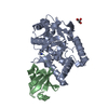
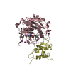
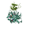
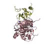
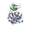
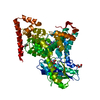
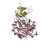
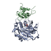
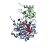
| Entry | Database: PDB / ID: 5lrx | |||||||||
|---|---|---|---|---|---|---|---|---|---|---|
| Title | Structure of A20 OTU domain bound to ubiquitin | |||||||||
 Components Components |
| |||||||||
 Keywords Keywords |  HYDROLASE / protease / deubiquitinase / OTU domain HYDROLASE / protease / deubiquitinase / OTU domain | |||||||||
| Function / homology |  Function and homology information Function and homology informationregulation of vascular wound healing / negative regulation of toll-like receptor 5 signaling pathway / negative regulation of nucleotide-binding oligomerization domain containing 1 signaling pathway / establishment of protein localization to vacuole / negative regulation of osteoclast proliferation / tolerance induction to lipopolysaccharide / negative regulation of CD40 signaling pathway / negative regulation of toll-like receptor 3 signaling pathway / negative regulation of B cell activation / negative regulation of nucleotide-binding oligomerization domain containing 2 signaling pathway ...regulation of vascular wound healing / negative regulation of toll-like receptor 5 signaling pathway / negative regulation of nucleotide-binding oligomerization domain containing 1 signaling pathway / establishment of protein localization to vacuole / negative regulation of osteoclast proliferation / tolerance induction to lipopolysaccharide / negative regulation of CD40 signaling pathway / negative regulation of toll-like receptor 3 signaling pathway / negative regulation of B cell activation / negative regulation of nucleotide-binding oligomerization domain containing 2 signaling pathway / negative regulation of toll-like receptor 2 signaling pathway / protein K11-linked deubiquitination / nucleotide-binding domain, leucine rich repeat containing receptor signaling pathway / negative regulation of chronic inflammatory response / negative regulation of toll-like receptor 4 signaling pathway / regulation of germinal center formation / protein K48-linked deubiquitination / B-1 B cell homeostasis / regulation of defense response to virus by host / regulation of tumor necrosis factor-mediated signaling pathway / Transferases; Acyltransferases; Aminoacyltransferases / protein K63-linked deubiquitination / positive regulation of hepatocyte proliferation / hypothalamus gonadotrophin-releasing hormone neuron development / negative regulation of bone resorption / female meiosis I / positive regulation of protein monoubiquitination / mitochondrion transport along microtubule / TNFR1-induced proapoptotic signaling / negative regulation of interleukin-1 beta production / fat pad development / negative regulation of interleukin-2 production / K63-linked polyubiquitin modification-dependent protein binding / K63-linked deubiquitinase activity / female gonad development / negative regulation of NF-kappaB transcription factor activity / seminiferous tubule development / protein deubiquitination / male meiosis I / positive regulation of intrinsic apoptotic signaling pathway by p53 class mediator / negative regulation of interleukin-6 production / response to muramyl dipeptide / negative regulation of tumor necrosis factor production / positive regulation of Wnt signaling pathway / protein K48-linked ubiquitination / negative regulation of endothelial cell apoptotic process / energy homeostasis / regulation of neuron apoptotic process / regulation of proteasomal protein catabolic process / negative regulation of canonical NF-kappaB signal transduction / negative regulation of extrinsic apoptotic signaling pathway via death domain receptors / Maturation of protein E / Maturation of protein E / ER Quality Control Compartment (ERQC) / Myoclonic epilepsy of Lafora / FLT3 signaling by CBL mutants / negative regulation of protein ubiquitination / Prevention of phagosomal-lysosomal fusion / IRAK2 mediated activation of TAK1 complex / Alpha-protein kinase 1 signaling pathway / Glycogen synthesis / IRAK1 recruits IKK complex / IRAK1 recruits IKK complex upon TLR7/8 or 9 stimulation / Membrane binding and targetting of GAG proteins / Endosomal Sorting Complex Required For Transport (ESCRT) / IRAK2 mediated activation of TAK1 complex upon TLR7/8 or 9 stimulation / PTK6 Regulates RTKs and Their Effectors AKT1 and DOK1 / Negative regulation of FLT3 / cytoskeleton organization / Constitutive Signaling by NOTCH1 HD Domain Mutants / Regulation of FZD by ubiquitination / TICAM1,TRAF6-dependent induction of TAK1 complex / NOTCH2 Activation and Transmission of Signal to the Nucleus / TICAM1-dependent activation of IRF3/IRF7 / APC/C:Cdc20 mediated degradation of Cyclin B / p75NTR recruits signalling complexes / Downregulation of ERBB4 signaling / TRAF6 mediated IRF7 activation in TLR7/8 or 9 signaling / APC-Cdc20 mediated degradation of Nek2A / PINK1-PRKN Mediated Mitophagy / TRAF6-mediated induction of TAK1 complex within TLR4 complex / Pexophagy / Regulation of innate immune responses to cytosolic DNA / VLDLR internalisation and degradation / InlA-mediated entry of Listeria monocytogenes into host cells / negative regulation of innate immune response / Downregulation of ERBB2:ERBB3 signaling / NF-kB is activated and signals survival / NRIF signals cell death from the nucleus / Regulation of PTEN localization / Activated NOTCH1 Transmits Signal to the Nucleus / Regulation of BACH1 activity / Translesion synthesis by REV1 / Synthesis of active ubiquitin: roles of E1 and E2 enzymes / Translesion synthesis by POLK / MAP3K8 (TPL2)-dependent MAPK1/3 activation / TICAM1, RIP1-mediated IKK complex recruitment / Downregulation of TGF-beta receptor signaling / Activation of IRF3, IRF7 mediated by TBK1, IKKε (IKBKE) / Translesion synthesis by POLISimilarity search - Function | |||||||||
| Biological species |  Homo sapiens (human) Homo sapiens (human) | |||||||||
| Method | X-RAY DIFFRACTION / SYNCHROTRON / MOLECULAR REPLACEMENT / Resolution: 2.85 Å | |||||||||
 Authors Authors | Mevissen, T.E.T. / Kulathu, Y. / Mulder, M.P.C. / Geurink, P.P. / Maslen, S.L. / Gersch, M. / Elliott, P.R. / Burke, J.E. / van Tol, B.D.M. / Akutsu, M. ...Mevissen, T.E.T. / Kulathu, Y. / Mulder, M.P.C. / Geurink, P.P. / Maslen, S.L. / Gersch, M. / Elliott, P.R. / Burke, J.E. / van Tol, B.D.M. / Akutsu, M. / El Oualid, F. / Kawasaki, M. / Freund, S.M.V. / Ovaa, H. / Komander, D. | |||||||||
| Funding support |  United Kingdom, 2items United Kingdom, 2items
| |||||||||
 Citation Citation | Journal: Nature / Year: 2016 Title: Molecular basis of Lys11-polyubiquitin specificity in the deubiquitinase Cezanne. Authors: Mevissen, T.E. / Kulathu, Y. / Mulder, M.P. / Geurink, P.P. / Maslen, S.L. / Gersch, M. / Elliott, P.R. / Burke, J.E. / van Tol, B.D. / Akutsu, M. / El Oualid, F. / Kawasaki, M. / Freund, S. ...Authors: Mevissen, T.E. / Kulathu, Y. / Mulder, M.P. / Geurink, P.P. / Maslen, S.L. / Gersch, M. / Elliott, P.R. / Burke, J.E. / van Tol, B.D. / Akutsu, M. / El Oualid, F. / Kawasaki, M. / Freund, S.M. / Ovaa, H. / Komander, D. | |||||||||
| History |
|
- Structure visualization
| Structure viewer | Molecule: MolmilJmol/JSmol |
|---|
- Downloads & links
-Download
| PDBx/mmCIF format | 5lrx.cif.gz | 590.9 KB | Display | PDBx/mmCIF format |
|---|---|---|---|---|
| PDB format | pdb5lrx.ent.gz | 500.6 KB | Display | PDB format |
| PDBx/mmJSON format | 5lrx.json.gz | Tree view | PDBx/mmJSON format | |
| Others |  Other downloads Other downloads |
-Validation report
| Arichive directory | https://data.pdbj.org/pub/pdb/validation_reports/lr/5lrxftp://data.pdbj.org/pub/pdb/validation_reports/lr/5lrx | HTTPS FTP |
|---|
-Related structure data


-Links
 PDBj
PDBj


- Assembly
| Deposited unit | 
| ||||||||
|---|---|---|---|---|---|---|---|---|---|
| 1 | 
| ||||||||
| 2 | 
| ||||||||
| 3 | 
| ||||||||
| 4 | 
| ||||||||
| Unit cell |
|
-Components
| #1: Protein | Mass: 43514.934 Da / Num. of mol.: 2 Source method: isolated from a genetically manipulated source Source: (gene. exp.) Homo sapiens (human) / Gene: TNFAIP3, OTUD7C / Production host:  Escherichia coli (E. coli) Escherichia coli (E. coli)References: UniProt: P21580, ubiquitinyl hydrolase 1, Ligases; Forming carbon-nitrogen bonds; Acid-amino-acid ligases (peptide synthases)#2: Protein | Mass: 8558.857 Da / Num. of mol.: 2 / Mutation: G76X Source method: isolated from a genetically manipulated source Details: Gly76 was replaced with propargylamide / Source: (gene. exp.) Homo sapiens (human) / Gene: UBB / Production host: Escherichia coli (E. coli) / References: UniProt: P0CG47#3: Protein | Mass: 43546.930 Da / Num. of mol.: 2 Source method: isolated from a genetically manipulated source Details: Cys103 is oxidized / Source: (gene. exp.) Homo sapiens (human) / Gene: TNFAIP3, OTUD7C / Production host: Escherichia coli (E. coli)References: UniProt: P21580, ubiquitinyl hydrolase 1, Ligases; Forming carbon-nitrogen bonds; Acid-amino-acid ligases (peptide synthases) |
|---|
-Experimental details
-Experiment
| Experiment | Method: X-RAY DIFFRACTION / Number of used crystals: 1 |
|---|
- Sample preparation
| Crystal | Density Matthews: 2.45 Å3/Da / Density % sol: 49.89 % |
|---|---|
| Crystal grow | Temperature: 287 K / Method: vapor diffusion, sitting drop Details: 0.1 M MES/imidazole (pH 6.5), 7% (w/v) PEG 8K, 20% ethylene glycol |
-Data collection
| Diffraction | Mean temperature: 100 K |
|---|---|
| Diffraction source | Source: SYNCHROTRON / Site: ESRF  / Beamline: ID29 / Wavelength: 0.96863 Å / Beamline: ID29 / Wavelength: 0.96863 Å |
| Detector | Type: DECTRIS PILATUS 6M / Detector: PIXEL / Date: Sep 16, 2012 |
| Radiation | Protocol: SINGLE WAVELENGTH / Monochromatic (M) / Laue (L): M / Scattering type: x-ray |
| Radiation wavelength | Wavelength: 0.96863 Å / Relative weight: 1 |
| Reflection | Resolution: 2.85→49.33 Å / Num. obs: 43448 / % possible obs: 99.4 % / Redundancy: 3.2 % / Rmerge(I) obs: 0.07 / Net I/σ(I): 9.6 |
| Reflection shell | Resolution: 2.85→2.96 Å / Redundancy: 3.3 % / Rmerge(I) obs: 0.384 / Mean I/σ(I) obs: 2.1 / % possible all: 99.8 |
- Processing
| Software |
| |||||||||||||||||||||||||||||||||||||||||||||||||||||||||||||||||||||||||||||||||||||||||||||||||||||||||||||||||||||||||||||||||||||||||||||||||||||||||||||||||||||||||||||||
|---|---|---|---|---|---|---|---|---|---|---|---|---|---|---|---|---|---|---|---|---|---|---|---|---|---|---|---|---|---|---|---|---|---|---|---|---|---|---|---|---|---|---|---|---|---|---|---|---|---|---|---|---|---|---|---|---|---|---|---|---|---|---|---|---|---|---|---|---|---|---|---|---|---|---|---|---|---|---|---|---|---|---|---|---|---|---|---|---|---|---|---|---|---|---|---|---|---|---|---|---|---|---|---|---|---|---|---|---|---|---|---|---|---|---|---|---|---|---|---|---|---|---|---|---|---|---|---|---|---|---|---|---|---|---|---|---|---|---|---|---|---|---|---|---|---|---|---|---|---|---|---|---|---|---|---|---|---|---|---|---|---|---|---|---|---|---|---|---|---|---|---|---|---|---|---|---|
| Refinement | Method to determine structure: MOLECULAR REPLACEMENT / Resolution: 2.85→49.325 Å / SU ML: 0.35 / Cross valid method: FREE R-VALUE / σ(F): 1.36 / Phase error: 26.15 / Stereochemistry target values: ML
| |||||||||||||||||||||||||||||||||||||||||||||||||||||||||||||||||||||||||||||||||||||||||||||||||||||||||||||||||||||||||||||||||||||||||||||||||||||||||||||||||||||||||||||||
| Solvent computation | Shrinkage radii: 0.9 Å / VDW probe radii: 1.11 Å / Solvent model: FLAT BULK SOLVENT MODEL | |||||||||||||||||||||||||||||||||||||||||||||||||||||||||||||||||||||||||||||||||||||||||||||||||||||||||||||||||||||||||||||||||||||||||||||||||||||||||||||||||||||||||||||||
| Refinement step | Cycle: LAST / Resolution: 2.85→49.325 Å
| |||||||||||||||||||||||||||||||||||||||||||||||||||||||||||||||||||||||||||||||||||||||||||||||||||||||||||||||||||||||||||||||||||||||||||||||||||||||||||||||||||||||||||||||
| Refine LS restraints |
| |||||||||||||||||||||||||||||||||||||||||||||||||||||||||||||||||||||||||||||||||||||||||||||||||||||||||||||||||||||||||||||||||||||||||||||||||||||||||||||||||||||||||||||||
| LS refinement shell |
| |||||||||||||||||||||||||||||||||||||||||||||||||||||||||||||||||||||||||||||||||||||||||||||||||||||||||||||||||||||||||||||||||||||||||||||||||||||||||||||||||||||||||||||||
| Refinement TLS params. | Method: refined / Refine-ID: X-RAY DIFFRACTION
| |||||||||||||||||||||||||||||||||||||||||||||||||||||||||||||||||||||||||||||||||||||||||||||||||||||||||||||||||||||||||||||||||||||||||||||||||||||||||||||||||||||||||||||||
| Refinement TLS group |
|
