 Movie
Movie Controller


+ Open data

- Basic information
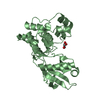
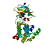
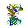
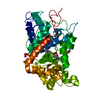
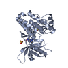
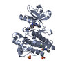
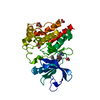
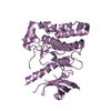
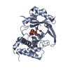
| Entry | Database: PDB / ID: 5ko1 | ||||||
|---|---|---|---|---|---|---|---|
| Title | Pseudokinase Domain of MLKL bound to Compound 4. | ||||||
 Components Components | Mixed lineage kinase domain-like protein | ||||||
 Keywords Keywords |  MEMBRANE PROTEINS/INHIBITOR / Pseudokinase domain MLKL Type1 inhibitor / MEMBRANE PROTEINS-INHIBITOR complex MEMBRANE PROTEINS/INHIBITOR / Pseudokinase domain MLKL Type1 inhibitor / MEMBRANE PROTEINS-INHIBITOR complex | ||||||
| Function / homology |  Function and homology information Function and homology informationexecution phase of necroptosis / Microbial modulation of RIPK1-mediated regulated necrosis / necroptotic signaling pathway / TRP channels / RIPK1-mediated regulated necrosis / protein homotrimerization / necroptotic process / Regulation of necroptotic cell death / cell junction / defense response to virus ...execution phase of necroptosis / Microbial modulation of RIPK1-mediated regulated necrosis / necroptotic signaling pathway / TRP channels / RIPK1-mediated regulated necrosis / protein homotrimerization / necroptotic process / Regulation of necroptotic cell death / cell junction / defense response to virus / cell surface receptor signaling pathway / protein-containing complex binding / protein kinase binding / ATP binding / identical protein binding / nucleus / plasma membrane / cytosol / cytoplasmSimilarity search - Function | ||||||
| Biological species |  Homo sapiens (human) Homo sapiens (human) | ||||||
| Method | X-RAY DIFFRACTION / SYNCHROTRON / MOLECULAR REPLACEMENT / Resolution: 2.16 Å | ||||||
 Authors Authors | Marcotte, D.J. | ||||||
 Citation Citation | Journal: Plos One / Year: 2016 Title: ATP-Competitive MLKL Binders Have No Functional Impact on Necroptosis. Authors: Ma, B. / Marcotte, D. / Paramasivam, M. / Michelsen, K. / Wang, T. / Bertolotti-Ciarlet, A. / Jones, J.H. / Moree, B. / Butko, M. / Salafsky, J. / Sun, X. / McKee, T. / Silvian, L.F. | ||||||
| History |
|
- Structure visualization
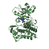
| Structure viewer | Molecule: MolmilJmol/JSmol |
|---|
- Downloads & links
-Download
| PDBx/mmCIF format | 5ko1.cif.gz | 72.7 KB | Display | PDBx/mmCIF format |
|---|---|---|---|---|
| PDB format | pdb5ko1.ent.gz | 52 KB | Display | PDB format |
| PDBx/mmJSON format | 5ko1.json.gz | Tree view | PDBx/mmJSON format | |
| Others |  Other downloads Other downloads |
-Validation report
| Arichive directory | https://data.pdbj.org/pub/pdb/validation_reports/ko/5ko1ftp://data.pdbj.org/pub/pdb/validation_reports/ko/5ko1 | HTTPS FTP |
|---|
-Related structure data
| Related structure data |  5knjC 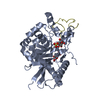 4wmiS C: citing same article ( S: Starting model for refinement |
|---|---|
| Similar structure data |


-Links
 PDBj
PDBj

- Assembly
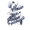
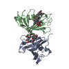
| Deposited unit | 
| ||||||||
|---|---|---|---|---|---|---|---|---|---|
| 1 |
| ||||||||
| Unit cell |
|
-Components
| #1: Protein | Mass: 32233.166 Da / Num. of mol.: 1 / Fragment: UNP residues 191-471 Source method: isolated from a genetically manipulated source Source: (gene. exp.) Homo sapiens (human) / Gene: MLKL / Production host:  Escherichia coli (E. coli) / References: UniProt: Q8NB16 Escherichia coli (E. coli) / References: UniProt: Q8NB16 |
|---|---|
| #2: Chemical | ChemComp-6UY / [(  Mass: 366.346 Da / Num. of mol.: 1 / Source method: obtained synthetically / Formula: C19H15FN4O3 Mass: 366.346 Da / Num. of mol.: 1 / Source method: obtained synthetically / Formula: C19H15FN4O3 |
| #3: Water | ChemComp-HOH / Water Mass: 18.015 Da / Num. of mol.: 130 / Source method: isolated from a natural source / Formula: H2O Mass: 18.015 Da / Num. of mol.: 130 / Source method: isolated from a natural source / Formula: H2O |
-Experimental details
-Experiment
| Experiment | Method: X-RAY DIFFRACTION / Number of used crystals: 1 |
|---|
- Sample preparation
| Crystal | Density Matthews: 2.62 Å3/Da / Density % sol: 53.13 % |
|---|---|
| Crystal grow | Temperature: 291 K / Method: vapor diffusion, sitting drop / pH: 6.5 / Details: 0.1M BisTRIS pH 6.5 20% PEG3350 |
-Data collection
| Diffraction | Mean temperature: 93 K | ||||||||||||||||||||||||||||||
|---|---|---|---|---|---|---|---|---|---|---|---|---|---|---|---|---|---|---|---|---|---|---|---|---|---|---|---|---|---|---|---|
| Diffraction source | Source: SYNCHROTRON / Site: APS  / Beamline: 31-ID / Wavelength: 0.98 Å / Beamline: 31-ID / Wavelength: 0.98 Å | ||||||||||||||||||||||||||||||
| Detector | Type: RAYONIX MX-225 / Detector: CCD / Date: Jul 22, 2015 | ||||||||||||||||||||||||||||||
| Radiation | Protocol: SINGLE WAVELENGTH / Monochromatic (M) / Laue (L): M / Scattering type: x-ray | ||||||||||||||||||||||||||||||
| Radiation wavelength | Wavelength: 0.98 Å / Relative weight: 1 | ||||||||||||||||||||||||||||||
| Reflection | Resolution: 2.16→63.53 Å / Num. obs: 18649 / % possible obs: 100 % / Redundancy: 7 % / CC1/2: 0.994 / Rmerge(I) obs: 0.143 / Rpim(I) all: 0.058 / Rrim(I) all: 0.155 / Net I/σ(I): 10 / Num. measured all: 131173 / Scaling rejects: 80 | ||||||||||||||||||||||||||||||
| Reflection shell | Diffraction-ID: 1 / Rejects: 0
|
- Processing
| Software |
| |||||||||||||||||||||||||||||||||||||||||||||||||||||||||||||||||||||||||||
|---|---|---|---|---|---|---|---|---|---|---|---|---|---|---|---|---|---|---|---|---|---|---|---|---|---|---|---|---|---|---|---|---|---|---|---|---|---|---|---|---|---|---|---|---|---|---|---|---|---|---|---|---|---|---|---|---|---|---|---|---|---|---|---|---|---|---|---|---|---|---|---|---|---|---|---|---|
| Refinement | Method to determine structure: MOLECULAR REPLACEMENT Starting model: 4WMI Resolution: 2.16→63.53 Å / Cor.coef. Fo:Fc: 0.944 / Cor.coef. Fo:Fc free: 0.927 / SU B: 5.554 / SU ML: 0.139 / Cross valid method: THROUGHOUT / σ(F): 0 / ESU R: 0.216 / ESU R Free: 0.18 / Stereochemistry target values: MAXIMUM LIKELIHOOD Details: HYDROGENS HAVE BEEN ADDED IN THE RIDING POSITIONS U VALUES : REFINED INDIVIDUALLY
| |||||||||||||||||||||||||||||||||||||||||||||||||||||||||||||||||||||||||||
| Solvent computation | Ion probe radii: 0.8 Å / Shrinkage radii: 0.8 Å / VDW probe radii: 1.2 Å / Solvent model: MASK | |||||||||||||||||||||||||||||||||||||||||||||||||||||||||||||||||||||||||||
| Displacement parameters | Biso max: 118.49 Å2 / Biso mean: 32.069 Å2 / Biso min: 14.38 Å2
| |||||||||||||||||||||||||||||||||||||||||||||||||||||||||||||||||||||||||||
| Refinement step | Cycle: final / Resolution: 2.16→63.53 Å
| |||||||||||||||||||||||||||||||||||||||||||||||||||||||||||||||||||||||||||
| Refine LS restraints |
| |||||||||||||||||||||||||||||||||||||||||||||||||||||||||||||||||||||||||||
| LS refinement shell | Resolution: 2.16→2.216 Å / Total num. of bins used: 20
|
