 Movie
Movie Controller


+ Open data

- Basic information
| Entry | Database: PDB / ID: 5ki9 | ||||||
|---|---|---|---|---|---|---|---|
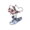
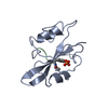
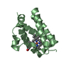
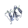
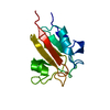
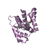
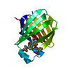
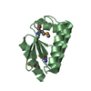
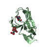
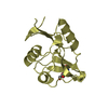
| Title | Crystal structure of human beta-defensin 4 (HBD4) | ||||||
 Components Components | Beta-defensin 104 | ||||||
 Keywords Keywords |  ANTIMICROBIAL PROTEIN / DEFENSIN / ANTIMICROBIAL / CHEMOTACTIC / DIMERIZATION ANTIMICROBIAL PROTEIN / DEFENSIN / ANTIMICROBIAL / CHEMOTACTIC / DIMERIZATION | ||||||
| Function / homology |  Function and homology informationBeta defensins / Defensins / cellular response to phorbol 13-acetate 12-myristate / positive chemotaxis / chemoattractant activity / monocyte chemotaxis / defense response to Gram-negative bacterium / defense response to Gram-positive bacterium / innate immune response / extracellular region Function and homology informationBeta defensins / Defensins / cellular response to phorbol 13-acetate 12-myristate / positive chemotaxis / chemoattractant activity / monocyte chemotaxis / defense response to Gram-negative bacterium / defense response to Gram-positive bacterium / innate immune response / extracellular regionSimilarity search - Function | ||||||
| Biological species |  Homo sapiens (human) Homo sapiens (human) | ||||||
| Method | X-RAY DIFFRACTION / SYNCHROTRON / MAD / Resolution: 1.6 Å | ||||||
 Authors Authors | Jacek, L. / Adam, P. / Marzenam, P. | ||||||
 Citation Citation | Journal: To Be Published Title: Human beta-Defensin 4: defensin without the "twist" Authors: Adam, P. / Marzenam, P. / Jerry, A. / Jacek, L. | ||||||
| History |
|
- Structure visualization
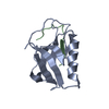
| Structure viewer | Molecule: MolmilJmol/JSmol |
|---|
- Downloads & links
-Download
| PDBx/mmCIF format | 5ki9.cif.gz | 30.6 KB | Display | PDBx/mmCIF format |
|---|---|---|---|---|
| PDB format | pdb5ki9.ent.gz | 22.8 KB | Display | PDB format |
| PDBx/mmJSON format | 5ki9.json.gz | Tree view | PDBx/mmJSON format | |
| Others |  Other downloads Other downloads |
-Validation report
| Arichive directory | https://data.pdbj.org/pub/pdb/validation_reports/ki/5ki9ftp://data.pdbj.org/pub/pdb/validation_reports/ki/5ki9 | HTTPS FTP |
|---|
-Related structure data
| Related structure data | |
|---|---|
| Similar structure data |
-Links
 PDBj
PDBj

- Assembly
| Deposited unit | 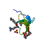
| ||||||||
|---|---|---|---|---|---|---|---|---|---|
| 1 | 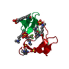
| ||||||||
| Unit cell |
| ||||||||
| Details | author states the biological assembly is unknown |
-Components
| #1: Protein/peptide | Mass: 5257.094 Da / Num. of mol.: 1 / Fragment: UNP residues 23-65 Source method: isolated from a genetically manipulated source Details: LYSINE RESIDUES ARE DI-METHYLATED / Source: (gene. exp.) Homo sapiens (human) / Gene: DEFB104A, DEFB104, DEFB4, DEFB104B / Production host:  Escherichia coli (E. coli) / References: UniProt: Q8WTQ1 Escherichia coli (E. coli) / References: UniProt: Q8WTQ1 | ||||
|---|---|---|---|---|---|
| #2: Chemical | Sulfate  Mass: 96.063 Da / Num. of mol.: 3 / Source method: obtained synthetically / Formula: SO4 Mass: 96.063 Da / Num. of mol.: 3 / Source method: obtained synthetically / Formula: SO4#3: Chemical | ChemComp-TFA / | Trifluoroacetic acid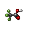  Mass: 114.023 Da / Num. of mol.: 1 / Source method: obtained synthetically / Formula: C2HF3O2 Mass: 114.023 Da / Num. of mol.: 1 / Source method: obtained synthetically / Formula: C2HF3O2#4: Water | ChemComp-HOH / | Water Mass: 18.015 Da / Num. of mol.: 17 / Source method: isolated from a natural source / Formula: H2O Mass: 18.015 Da / Num. of mol.: 17 / Source method: isolated from a natural source / Formula: H2O |
-Experimental details
-Experiment
| Experiment | Method: X-RAY DIFFRACTION / Number of used crystals: 1 |
|---|
- Sample preparation
| Crystal | Density Matthews: 2.19 Å3/Da / Density % sol: 43.82 % |
|---|---|
| Crystal grow | Temperature: 293 K / Method: vapor diffusion, hanging drop / pH: 4.5 Details: 25.5% PEG 8,000, 0.085 M sodium acetate buffer pH 4.5, 0.17 M Lithium Sulfate, 15% Glycerol |
-Data collection
| Diffraction | Mean temperature: 100 K | |||||||||||||||||||||||||||||||||||||||||||||||||||||||||||||||||||||||||||||||||||||||||||||||||||||||||
|---|---|---|---|---|---|---|---|---|---|---|---|---|---|---|---|---|---|---|---|---|---|---|---|---|---|---|---|---|---|---|---|---|---|---|---|---|---|---|---|---|---|---|---|---|---|---|---|---|---|---|---|---|---|---|---|---|---|---|---|---|---|---|---|---|---|---|---|---|---|---|---|---|---|---|---|---|---|---|---|---|---|---|---|---|---|---|---|---|---|---|---|---|---|---|---|---|---|---|---|---|---|---|---|---|---|---|
| Diffraction source | Source: SYNCHROTRON / Site: APS  / Beamline: 22-ID / Wavelength: 1 Å / Beamline: 22-ID / Wavelength: 1 Å | |||||||||||||||||||||||||||||||||||||||||||||||||||||||||||||||||||||||||||||||||||||||||||||||||||||||||
| Detector | Type: MARMOSAIC 300 mm CCD / Detector: CCD / Date: Oct 10, 2010 | |||||||||||||||||||||||||||||||||||||||||||||||||||||||||||||||||||||||||||||||||||||||||||||||||||||||||
| Radiation | Protocol: SINGLE WAVELENGTH / Monochromatic (M) / Laue (L): M / Scattering type: x-ray | |||||||||||||||||||||||||||||||||||||||||||||||||||||||||||||||||||||||||||||||||||||||||||||||||||||||||
| Radiation wavelength | Wavelength: 1 Å / Relative weight: 1 | |||||||||||||||||||||||||||||||||||||||||||||||||||||||||||||||||||||||||||||||||||||||||||||||||||||||||
| Reflection | Resolution: 1.55→50 Å / Num. obs: 6994 / % possible obs: 95.6 % / Redundancy: 16.6 % / Rmerge(I) obs: 0.059 / Rpim(I) all: 0.014 / Rrim(I) all: 0.06 / Χ2: 1.697 / Net I/av σ(I): 90.17 / Net I/σ(I): 16.7 / Num. measured all: 116371 | |||||||||||||||||||||||||||||||||||||||||||||||||||||||||||||||||||||||||||||||||||||||||||||||||||||||||
| Reflection shell |
|
-Phasing
| Phasing | Method: MAD |
|---|
- Processing
| Software |
| ||||||||||||||||||||||||||||||||||||||||||||||||||||||||||||||||||||||
|---|---|---|---|---|---|---|---|---|---|---|---|---|---|---|---|---|---|---|---|---|---|---|---|---|---|---|---|---|---|---|---|---|---|---|---|---|---|---|---|---|---|---|---|---|---|---|---|---|---|---|---|---|---|---|---|---|---|---|---|---|---|---|---|---|---|---|---|---|---|---|---|
| Refinement | Method to determine structure: MAD / Resolution: 1.6→15 Å / Cor.coef. Fo:Fc: 0.956 / Cor.coef. Fo:Fc free: 0.931 / SU B: 5.658 / SU ML: 0.087 / SU R Cruickshank DPI: 0.1588 / Cross valid method: THROUGHOUT / σ(F): 0 / ESU R: 0.159 / ESU R Free: 0.115 Details: HYDROGENS HAVE BEEN ADDED IN THE RIDING POSITIONS U VALUES : REFINED INDIVIDUALLY
| ||||||||||||||||||||||||||||||||||||||||||||||||||||||||||||||||||||||
| Solvent computation | Ion probe radii: 0.8 Å / Shrinkage radii: 0.8 Å / VDW probe radii: 1.2 Å | ||||||||||||||||||||||||||||||||||||||||||||||||||||||||||||||||||||||
| Displacement parameters | Biso max: 170.24 Å2 / Biso mean: 51.173 Å2 / Biso min: 30.09 Å2
| ||||||||||||||||||||||||||||||||||||||||||||||||||||||||||||||||||||||
| Refinement step | Cycle: final / Resolution: 1.6→15 Å
| ||||||||||||||||||||||||||||||||||||||||||||||||||||||||||||||||||||||
| Refine LS restraints |
| ||||||||||||||||||||||||||||||||||||||||||||||||||||||||||||||||||||||
| LS refinement shell | Resolution: 1.6→1.641 Å / Total num. of bins used: 20
|
