 Movie
Movie Controller

[English] 日本語
 Yorodumi
- PDB-5k39: THE TYPE II COHESIN DOCKERIN COMPLEX FROM CLOSTRIDIUM THERMOCELLUM -
+ Open data

- Basic information
| Entry | Database: PDB / ID: 5k39 | ||||||
|---|---|---|---|---|---|---|---|
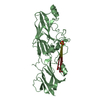
| Title | THE TYPE II COHESIN DOCKERIN COMPLEX FROM CLOSTRIDIUM THERMOCELLUM | ||||||
 Components Components |
| ||||||
 Keywords Keywords |  STRUCTURAL PROTEIN / S-LAYER / SECRETED / CELL WALL / MEMBRANE PROTEIN STRUCTURAL PROTEIN / S-LAYER / SECRETED / CELL WALL / MEMBRANE PROTEIN | ||||||
| Function / homology |  Function and homology informationS-layer / cellulose catabolic process / polysaccharide catabolic process / hydrolase activity, hydrolyzing O-glycosyl compounds / carbohydrate binding / extracellular region / metal ion binding Function and homology informationS-layer / cellulose catabolic process / polysaccharide catabolic process / hydrolase activity, hydrolyzing O-glycosyl compounds / carbohydrate binding / extracellular region / metal ion bindingSimilarity search - Function | ||||||
| Biological species |  Ruminiclostridium thermocellum 27405 (bacteria)Clostridium thermocellum 27405 (bacteria) Ruminiclostridium thermocellum 27405 (bacteria)Clostridium thermocellum 27405 (bacteria) | ||||||
| Method | X-RAY DIFFRACTION / SYNCHROTRON / MOLECULAR REPLACEMENT / Resolution: 1.98 Å | ||||||
 Authors Authors | Viegas, A. / Pinheiro, B. / Bras, J.L.A. / Romao, M.J. / Alves, V. / Carvalho, A.L. / Fontes, C.M.G.A. | ||||||
 Citation Citation | Journal: Sci Rep / Year: 2016 Title: Diverse specificity of cellulosome attachment to the bacterial cell surface. Authors: Bras, J.L. / Pinheiro, B.A. / Cameron, K. / Cuskin, F. / Viegas, A. / Najmudin, S. / Bule, P. / Pires, V.M. / Romao, M.J. / Bayer, E.A. / Spencer, H.L. / Smith, S. / Gilbert, H.J. / Alves, V. ...Authors: Bras, J.L. / Pinheiro, B.A. / Cameron, K. / Cuskin, F. / Viegas, A. / Najmudin, S. / Bule, P. / Pires, V.M. / Romao, M.J. / Bayer, E.A. / Spencer, H.L. / Smith, S. / Gilbert, H.J. / Alves, V.D. / Carvalho, A.L. / Fontes, C.M. | ||||||
| History |
| ||||||
| Remark 700 | SHEET THE SHEET STRUCTURE OF THIS MOLECULE IS BIFURCATED. IN ORDER TO REPRESENT THIS FEATURE IN THE ...SHEET THE SHEET STRUCTURE OF THIS MOLECULE IS BIFURCATED. IN ORDER TO REPRESENT THIS FEATURE IN THE SHEET RECORDS BELOW, TWO SHEETS ARE DEFINED. |
- Structure visualization
| Structure viewer | Molecule: MolmilJmol/JSmol |
|---|
- Downloads & links
-Download
| PDBx/mmCIF format | 5k39.cif.gz | 85.3 KB | Display | PDBx/mmCIF format |
|---|---|---|---|---|
| PDB format | pdb5k39.ent.gz | 62.2 KB | Display | PDB format |
| PDBx/mmJSON format | 5k39.json.gz | Tree view | PDBx/mmJSON format | |
| Others |  Other downloads Other downloads |
-Validation report
| Arichive directory | https://data.pdbj.org/pub/pdb/validation_reports/k3/5k39ftp://data.pdbj.org/pub/pdb/validation_reports/k3/5k39 | HTTPS FTP |
|---|
-Related structure data
| Related structure data |  5g5dC  5m0yC  2b59S  2mb3S S: Starting model for refinement C: citing same article ( |
|---|---|
| Similar structure data |


-Links
 PDBj
PDBj
- Assembly
| Deposited unit | 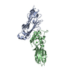
| ||||||||
|---|---|---|---|---|---|---|---|---|---|
| 1 |
| ||||||||
| Unit cell |
| ||||||||
| Components on special symmetry positions |
|
-Components
| #1: Protein | Mass: 18947.541 Da / Num. of mol.: 1 / Fragment: COHESION 2 DOMAIN, RESIDUES 205-364 Source method: isolated from a genetically manipulated source Source: (gene. exp.) Ruminiclostridium thermocellum 27405 (bacteria)Gene: AD2_00639 / Plasmid: pCF1 / Details (production host): altered pET21 plasmid / Production host: Escherichia coli BL21 (bacteria) / References: UniProt: A0A0U3TQ90, UniProt: Q06853*PLUS | ||
|---|---|---|---|
| #2: Protein | Mass: 18245.408 Da / Num. of mol.: 1 / Fragment: RESIDUES 2015-2177 Source method: isolated from a genetically manipulated source Source: (gene. exp.) Clostridium thermocellum 27405 (bacteria)Gene: Cthe_1806 / Production host: Escherichia coli (E. coli) / References: UniProt: A3DGE8 | ||
| #3: Chemical |   Mass: 40.078 Da / Num. of mol.: 2 / Source method: obtained synthetically / Formula: Ca Mass: 40.078 Da / Num. of mol.: 2 / Source method: obtained synthetically / Formula: Ca#4: Water | ChemComp-HOH / | Water Mass: 18.015 Da / Num. of mol.: 322 / Source method: isolated from a natural source / Formula: H2O Mass: 18.015 Da / Num. of mol.: 322 / Source method: isolated from a natural source / Formula: H2O |
-Experimental details
-Experiment
| Experiment | Method: X-RAY DIFFRACTION / Number of used crystals: 1 |
|---|
- Sample preparation
| Crystal | Density Matthews: 2.2 Å3/Da / Density % sol: 43.13 % |
|---|---|
| Crystal grow | Temperature: 293 K / Method: vapor diffusion, hanging drop Details: 12% (m/v) polyethyleneglycol (PEG) 3350, 4% tacsimate, pH 5.0 |
-Data collection
| Diffraction | Mean temperature: 100 K |
|---|---|
| Diffraction source | Source: SYNCHROTRON / Site: ESRF  / Beamline: ID14-4 / Wavelength: 0.9735 Å / Beamline: ID14-4 / Wavelength: 0.9735 Å |
| Detector | Type: ADSC QUANTUM 315r / Detector: CCD / Date: Feb 28, 2008 |
| Radiation | Protocol: SINGLE WAVELENGTH / Monochromatic (M) / Laue (L): M / Scattering type: x-ray |
| Radiation wavelength | Wavelength: 0.9735 Å / Relative weight: 1 |
| Reflection | Resolution: 1.98→39.31 Å / Num. obs: 21909 / % possible obs: 97.8 % / Observed criterion σ(I): 4 / Redundancy: 3.6 % / Rmerge(I) obs: 0.09 / Net I/σ(I): 13.1 |
| Reflection shell | Resolution: 1.98→2.09 Å / Redundancy: 3.7 % / Rmerge(I) obs: 0.17 / Mean I/σ(I) obs: 5.5 / % possible all: 98.2 |
- Processing
| Software |
| ||||||||||||||||||||||||||||||||||||||||||||||||||||||||||||||||||||||||||||||||||||||||||||||||||||||||||||||||||||||||||||||||||||||||||||||||||||||||||||||||||||||||||||||||||||||
|---|---|---|---|---|---|---|---|---|---|---|---|---|---|---|---|---|---|---|---|---|---|---|---|---|---|---|---|---|---|---|---|---|---|---|---|---|---|---|---|---|---|---|---|---|---|---|---|---|---|---|---|---|---|---|---|---|---|---|---|---|---|---|---|---|---|---|---|---|---|---|---|---|---|---|---|---|---|---|---|---|---|---|---|---|---|---|---|---|---|---|---|---|---|---|---|---|---|---|---|---|---|---|---|---|---|---|---|---|---|---|---|---|---|---|---|---|---|---|---|---|---|---|---|---|---|---|---|---|---|---|---|---|---|---|---|---|---|---|---|---|---|---|---|---|---|---|---|---|---|---|---|---|---|---|---|---|---|---|---|---|---|---|---|---|---|---|---|---|---|---|---|---|---|---|---|---|---|---|---|---|---|---|---|
| Refinement | Method to determine structure: MOLECULAR REPLACEMENT Starting model: PDB ENTRIES 2MB3 AND 2B59 Resolution: 1.98→18.83 Å / Cor.coef. Fo:Fc: 0.945 / Cor.coef. Fo:Fc free: 0.906 / SU B: 7.842 / SU ML: 0.124 / Cross valid method: THROUGHOUT / ESU R: 0.225 / ESU R Free: 0.194 / Details: HYDROGENS HAVE BEEN ADDED IN THE RIDING POSITIONS.
| ||||||||||||||||||||||||||||||||||||||||||||||||||||||||||||||||||||||||||||||||||||||||||||||||||||||||||||||||||||||||||||||||||||||||||||||||||||||||||||||||||||||||||||||||||||||
| Solvent computation | Ion probe radii: 0.8 Å / Shrinkage radii: 0.8 Å / VDW probe radii: 1.2 Å | ||||||||||||||||||||||||||||||||||||||||||||||||||||||||||||||||||||||||||||||||||||||||||||||||||||||||||||||||||||||||||||||||||||||||||||||||||||||||||||||||||||||||||||||||||||||
| Displacement parameters | Biso mean: 20.01 Å2
| ||||||||||||||||||||||||||||||||||||||||||||||||||||||||||||||||||||||||||||||||||||||||||||||||||||||||||||||||||||||||||||||||||||||||||||||||||||||||||||||||||||||||||||||||||||||
| Refinement step | Cycle: LAST / Resolution: 1.98→18.83 Å
| ||||||||||||||||||||||||||||||||||||||||||||||||||||||||||||||||||||||||||||||||||||||||||||||||||||||||||||||||||||||||||||||||||||||||||||||||||||||||||||||||||||||||||||||||||||||
| Refine LS restraints |
|