 Movie
Movie Controller

[English] 日本語
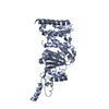
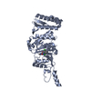
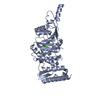
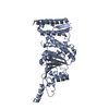
 Yorodumi
- PDB-5dtr: Crystal structure of Dot1L in complex with inhibitor CPD5 [N-(2,6... -
+ Open data

- Basic information
| Entry | Database: PDB / ID: 5dtr | ||||||
|---|---|---|---|---|---|---|---|
| Title | Crystal structure of Dot1L in complex with inhibitor CPD5 [N-(2,6-dichlorophenyl)-4-methoxy-N-methylquinolin-6-amine] | ||||||
 Components Components | Histone-lysine N-methyltransferase, H3 lysine-79 specific Histone methyltransferase Histone methyltransferase | ||||||
 Keywords Keywords | TRANSFERASE / Inhibitor / Complex / Methyltransferase | ||||||
| Function / homology |  Function and homology information Function and homology informationhistone H3K79 trimethyltransferase activity / [histone H3]-lysine79 N-trimethyltransferase / histone H3K79 methyltransferase activity / regulation of transcription regulatory region DNA binding / histone H3 methyltransferase activity / regulation of receptor signaling pathway via JAK-STAT / histone methyltransferase activity / heterochromatin formation / telomere organization / DNA damage checkpoint signaling ...histone H3K79 trimethyltransferase activity / [histone H3]-lysine79 N-trimethyltransferase / histone H3K79 methyltransferase activity / regulation of transcription regulatory region DNA binding / histone H3 methyltransferase activity / regulation of receptor signaling pathway via JAK-STAT / histone methyltransferase activity / heterochromatin formation / telomere organization / DNA damage checkpoint signaling / PKMTs methylate histone lysines / gene expression / methylation / RNA polymerase II-specific DNA-binding transcription factor binding / nucleic acid binding / transcription coactivator activity / intracellular membrane-bounded organelle / DNA repair / positive regulation of cell population proliferation / positive regulation of transcription by RNA polymerase II / protein-containing complex / DNA binding / nucleoplasm / nucleus / cytoplasmSimilarity search - Function | ||||||
| Biological species |  Homo sapiens (human) Homo sapiens (human) | ||||||
| Method | X-RAY DIFFRACTION / SYNCHROTRON / MOLECULAR REPLACEMENT / Resolution: 2.34 Å | ||||||
 Authors Authors | Scheufler, C. / Be, C. / Moebitz, H. / Stauffer, F. | ||||||
 Citation Citation | Journal: Acs Med.Chem.Lett. / Year: 2016 Title: Optimization of a Fragment-Based Screening Hit toward Potent DOT1L Inhibitors Interacting in an Induced Binding Pocket. Authors: Scheufler, C. / Mobitz, H. / Gaul, C. / Ragot, C. / Be, C. / Fernandez, C. / Beyer, K.S. / Tiedt, R. / Stauffer, F. | ||||||
| History |
|
- Structure visualization
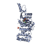
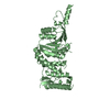
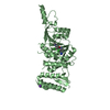
| Structure viewer | Molecule: MolmilJmol/JSmol |
|---|
- Downloads & links
-Download
| PDBx/mmCIF format | 5dtr.cif.gz | 275.2 KB | Display | PDBx/mmCIF format |
|---|---|---|---|---|
| PDB format | pdb5dtr.ent.gz | 222.2 KB | Display | PDB format |
| PDBx/mmJSON format | 5dtr.json.gz | Tree view | PDBx/mmJSON format | |
| Others |  Other downloads Other downloads |
-Validation report
| Arichive directory | https://data.pdbj.org/pub/pdb/validation_reports/dt/5dtrftp://data.pdbj.org/pub/pdb/validation_reports/dt/5dtr | HTTPS FTP |
|---|
-Related structure data
| Related structure data |  5dtmC  5dtqC 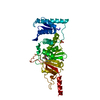 1nw3S S: Starting model for refinement C: citing same article ( |
|---|---|
| Similar structure data |


-Links
 PDBj
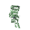
PDBj- Assembly
| Deposited unit | 
| ||||||||
|---|---|---|---|---|---|---|---|---|---|
| 1 | 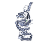
| ||||||||
| 2 | 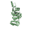
| ||||||||
| Unit cell |
|
-Components
| #1: Protein | Histone methyltransferase / DOT1-like protein / Histone H3-K79 methyltransferase / H3-K79-HMTase / Lysine N-methyltransferase 4 Mass: 38456.594 Da / Num. of mol.: 2 Source method: isolated from a genetically manipulated source Source: (gene. exp.) Homo sapiens (human) / Gene: DOT1L, KIAA1814, KMT4 / Production host:  Escherichia coli (E. coli) Escherichia coli (E. coli)References: UniProt: Q8TEK3, histone-lysine N-methyltransferase#2: Chemical |   Mass: 333.212 Da / Num. of mol.: 2 / Source method: obtained synthetically / Formula: C17H14Cl2N2O Mass: 333.212 Da / Num. of mol.: 2 / Source method: obtained synthetically / Formula: C17H14Cl2N2O#3: Water | ChemComp-HOH / | Water Mass: 18.015 Da / Num. of mol.: 315 / Source method: isolated from a natural source / Formula: H2O Mass: 18.015 Da / Num. of mol.: 315 / Source method: isolated from a natural source / Formula: H2O |
|---|
-Experimental details
-Experiment
| Experiment | Method: X-RAY DIFFRACTION |
|---|
- Sample preparation
| Crystal | Density Matthews: 3.48 Å3/Da / Density % sol: 64.69 % |
|---|---|
| Crystal grow | Temperature: 293 K / Method: vapor diffusion / Details: 1.2M K/Na tartrate, 0.1M Hepes pH6.6 |
-Data collection
| Diffraction | Mean temperature: 100 K |
|---|---|
| Diffraction source | Source: SYNCHROTRON / Site: SLS  / Beamline: X10SA / Wavelength: 0.99985 Å / Beamline: X10SA / Wavelength: 0.99985 Å |
| Detector | Type: DECTRIS PILATUS 6M / Detector: PIXEL / Date: May 18, 2012 |
| Radiation | Protocol: SINGLE WAVELENGTH / Monochromatic (M) / Laue (L): M / Scattering type: x-ray |
| Radiation wavelength | Wavelength: 0.99985 Å / Relative weight: 1 |
| Reflection | Resolution: 2.34→50 Å / Num. all: 44848 / Num. obs: 44838 / % possible obs: 100 % / Redundancy: 10.2 % / Biso Wilson estimate: 49.04 Å2 / Rmerge(I) obs: 0.087 / Net I/σ(I): 21.31 |
| Reflection shell | Resolution: 2.34→2.4 Å / Redundancy: 10.3 % / Rmerge(I) obs: 0.925 / Mean I/σ(I) obs: 3.25 / % possible all: 100 |
- Processing
| Software |
| ||||||||||||||||||||||||||||||||||||||||||||||||||||||||||||||||||||||||||||||||||||||||||||||||||||||||||||||||||
|---|---|---|---|---|---|---|---|---|---|---|---|---|---|---|---|---|---|---|---|---|---|---|---|---|---|---|---|---|---|---|---|---|---|---|---|---|---|---|---|---|---|---|---|---|---|---|---|---|---|---|---|---|---|---|---|---|---|---|---|---|---|---|---|---|---|---|---|---|---|---|---|---|---|---|---|---|---|---|---|---|---|---|---|---|---|---|---|---|---|---|---|---|---|---|---|---|---|---|---|---|---|---|---|---|---|---|---|---|---|---|---|---|---|---|---|
| Refinement | Method to determine structure: MOLECULAR REPLACEMENT Starting model: 1nw3 Resolution: 2.34→42.46 Å / Cor.coef. Fo:Fc: 0.9442 / Cor.coef. Fo:Fc free: 0.9223 / SU R Cruickshank DPI: 0.195 / Cross valid method: THROUGHOUT / σ(F): 0 / SU R Blow DPI: 0.196 / SU Rfree Blow DPI: 0.17 / SU Rfree Cruickshank DPI: 0.171
| ||||||||||||||||||||||||||||||||||||||||||||||||||||||||||||||||||||||||||||||||||||||||||||||||||||||||||||||||||
| Displacement parameters | Biso mean: 56.64 Å2
| ||||||||||||||||||||||||||||||||||||||||||||||||||||||||||||||||||||||||||||||||||||||||||||||||||||||||||||||||||
| Refine analyze | Luzzati coordinate error obs: 0.305 Å | ||||||||||||||||||||||||||||||||||||||||||||||||||||||||||||||||||||||||||||||||||||||||||||||||||||||||||||||||||
| Refinement step | Cycle: 1 / Resolution: 2.34→42.46 Å
| ||||||||||||||||||||||||||||||||||||||||||||||||||||||||||||||||||||||||||||||||||||||||||||||||||||||||||||||||||
| Refine LS restraints |
| ||||||||||||||||||||||||||||||||||||||||||||||||||||||||||||||||||||||||||||||||||||||||||||||||||||||||||||||||||
| LS refinement shell | Resolution: 2.34→2.4 Å / Total num. of bins used: 20
| ||||||||||||||||||||||||||||||||||||||||||||||||||||||||||||||||||||||||||||||||||||||||||||||||||||||||||||||||||
| Refinement TLS params. | Method: refined / Refine-ID: X-RAY DIFFRACTION
| ||||||||||||||||||||||||||||||||||||||||||||||||||||||||||||||||||||||||||||||||||||||||||||||||||||||||||||||||||
| Refinement TLS group |
|
