 Movie
Movie Controller

[English] 日本語
 Yorodumi
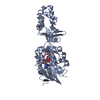
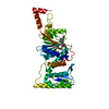
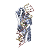
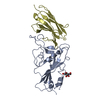
- PDB-3um3: Crystal structure of the Brox Bro1 domain in complex with the C-t... -
+ Open data

- Basic information
| Entry | Database: PDB / ID: 3um3 | ||||||
|---|---|---|---|---|---|---|---|
| Title | Crystal structure of the Brox Bro1 domain in complex with the C-terminal tail of CHMP4B | ||||||
 Components Components |
| ||||||
 Keywords Keywords |  MEMBRANE PROTEIN/TRANSPORT PROTEIN / alpha-helix of C-terminal tail of CHMP4B / ESCRT-III / CHMPs / MEMBRANE PROTEIN-TRANSPORT PROTEIN complex / BROX MEMBRANE PROTEIN/TRANSPORT PROTEIN / alpha-helix of C-terminal tail of CHMP4B / ESCRT-III / CHMPs / MEMBRANE PROTEIN-TRANSPORT PROTEIN complex / BROX | ||||||
| Function / homology |  Function and homology information Function and homology informationubiquitin-independent protein catabolic process via the multivesicular body sorting pathway / maintenance of lens transparency / amphisome membrane / multivesicular body-lysosome fusion / viral budding / vesicle fusion with vacuole / late endosome to lysosome transport / ESCRT III complex / kinetochore microtubule / late endosome to vacuole transport via multivesicular body sorting pathway ...ubiquitin-independent protein catabolic process via the multivesicular body sorting pathway / maintenance of lens transparency / amphisome membrane / multivesicular body-lysosome fusion / viral budding / vesicle fusion with vacuole / late endosome to lysosome transport / ESCRT III complex / kinetochore microtubule / late endosome to vacuole transport via multivesicular body sorting pathway / mitotic nuclear membrane reassembly / regulation of centrosome duplication / nuclear membrane reassembly / Sealing of the nuclear envelope (NE) by ESCRT-III / midbody abscission / post-translational protein targeting to endoplasmic reticulum membrane / multivesicular body sorting pathway / vesicle budding from membrane / membrane fission / plasma membrane repair / membrane coat / multivesicular body membrane / ubiquitin-dependent protein catabolic process via the multivesicular body sorting pathway / exit from mitosis / multivesicular body assembly / regulation of mitotic spindle assembly / nervous system process / Translation of Replicase and Assembly of the Replication Transcription Complex / nucleus organization / Macroautophagy / mitotic metaphase chromosome alignment / viral budding via host ESCRT complex / autophagosome maturation / autophagosome membrane / mitotic cytokinesis / protein polymerization / Pyroptosis / nuclear pore / Endosomal Sorting Complex Required For Transport (ESCRT) / multivesicular body / viral budding from plasma membrane / HCMV Late Events / regulation of autophagy / macroautophagy / Late endosomal microautophagy / Budding and maturation of HIV virion / cytoplasmic side of plasma membrane / kinetochore / autophagy / protein transport / nuclear envelope / Translation of Replicase and Assembly of the Replication Transcription Complex / midbody / nuclear membrane / vesicle / endosome / cadherin binding / lysosomal membrane / protein homodimerization activity / extracellular exosome / identical protein binding / nucleus / plasma membrane / cytosol / cytoplasmSimilarity search - Function | ||||||
| Biological species |  Homo sapiens (human) Homo sapiens (human) | ||||||
| Method | X-RAY DIFFRACTION / SYNCHROTRON / MOLECULAR REPLACEMENT / Resolution: 3.8 Å | ||||||
 Authors Authors | Jiang, J.S. / Mu, R.L. / Xiao, T. | ||||||
 Citation Citation | Journal: Structure / Year: 2012 Title: Two Distinct Binding Modes Define the Interaction of Brox with the C-Terminal Tails of CHMP5 and CHMP4B. Authors: Mu, R. / Dussupt, V. / Jiang, J. / Sette, P. / Rudd, V. / Chuenchor, W. / Bello, N.F. / Bouamr, F. / Xiao, T.S. | ||||||
| History |
|
- Structure visualization
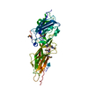
| Structure viewer | Molecule: MolmilJmol/JSmol |
|---|
- Downloads & links
-Download
| PDBx/mmCIF format | 3um3.cif.gz | 85.2 KB | Display | PDBx/mmCIF format |
|---|---|---|---|---|
| PDB format | pdb3um3.ent.gz | 64.6 KB | Display | PDB format |
| PDBx/mmJSON format | 3um3.json.gz | Tree view | PDBx/mmJSON format | |
| Others |  Other downloads Other downloads |
-Validation report
| Arichive directory | https://data.pdbj.org/pub/pdb/validation_reports/um/3um3ftp://data.pdbj.org/pub/pdb/validation_reports/um/3um3 | HTTPS FTP |
|---|
-Related structure data
| Related structure data | 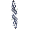 3ulyC 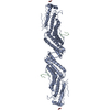 3um0C 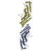 3um1C 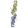 3um2C  3r9mS S: Starting model for refinement C: citing same article ( |
|---|---|
| Similar structure data |


-Links
 PDBj
PDBj


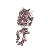
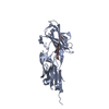
- Assembly
| Deposited unit | 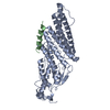
| ||||||||
|---|---|---|---|---|---|---|---|---|---|
| 1 |
| ||||||||
| Unit cell |
|
-Components
| #1: Protein | Mass: 46521.863 Da / Num. of mol.: 1 / Fragment: Brox bro1 domain 2-377 Source method: isolated from a genetically manipulated source Source: (gene. exp.) Homo sapiens (human) / Gene: BROFTI, BROX, C1orf58 / Plasmid: pET30a / Production host:  Escherichia coli (E. coli) / Strain (production host): BL21(DE3) / References: UniProt: Q5VW32 Escherichia coli (E. coli) / Strain (production host): BL21(DE3) / References: UniProt: Q5VW32 |
|---|---|
| #2: Protein | Mass: 11699.792 Da / Num. of mol.: 1 / Fragment: C-terminal tail of CHMP4B 121-224 Source method: isolated from a genetically manipulated source Source: (gene. exp.) Homo sapiens (human) / Gene: C20orf178, CHMP4B, SHAX1 / Plasmid: pET30a / Production host: Escherichia coli (E. coli) / Strain (production host): BL21(DE3) / References: UniProt: Q9H444 |
-Experimental details
-Experiment
| Experiment | Method: X-RAY DIFFRACTION / Number of used crystals: 1 |
|---|
- Sample preparation
| Crystal grow | Temperature: 273 K / Method: vapor diffusion, hanging drop / pH: 7.2 Details: 1.0M sodium citrate, 0.1M imidazol, 15% glycerol, pH 7.2, VAPOR DIFFUSION, HANGING DROP, temperature 273K |
|---|
-Data collection
| Diffraction | Mean temperature: 100 K |
|---|---|
| Diffraction source | Source: SYNCHROTRON / Site: NSLS  / Beamline: X29A / Wavelength: 1.075 Å / Beamline: X29A / Wavelength: 1.075 Å |
| Detector | Type: ADSC QUANTUM 315 / Detector: CCD / Date: Apr 16, 2010 |
| Radiation | Protocol: SINGLE WAVELENGTH / Monochromatic (M) / Laue (L): M / Scattering type: x-ray |
| Radiation wavelength | Wavelength: 1.075 Å / Relative weight: 1 |
| Reflection | Resolution: 3.8→50 Å / Num. obs: 21693 / % possible obs: 99.3 % / Observed criterion σ(F): 0 / Observed criterion σ(I): 0 / Redundancy: 4.3 % / Biso Wilson estimate: 145 Å2 / Rmerge(I) obs: 0.036 / Net I/σ(I): 18.3 |
- Processing
| Software |
| |||||||||||||||||||||||||
|---|---|---|---|---|---|---|---|---|---|---|---|---|---|---|---|---|---|---|---|---|---|---|---|---|---|---|
| Refinement | Method to determine structure: MOLECULAR REPLACEMENT Starting model: PDB ENTRY 3R9M Resolution: 3.8→48.76 Å / Rfactor Rfree error: 0.005 / Data cutoff high absF: 5336689.33 / Data cutoff low absF: 0 / Isotropic thermal model: RESTRAINED / Cross valid method: THROUGHOUT / σ(F): 0 / Stereochemistry target values: Engh & Huber / Details: BULK SOLVENT MODEL USED
| |||||||||||||||||||||||||
| Solvent computation | Solvent model: FLAT MODEL / Bsol: 42.2265 Å2 / ksol: 0.33 e/Å3 | |||||||||||||||||||||||||
| Displacement parameters | Biso mean: 83.3 Å2
| |||||||||||||||||||||||||
| Refine analyze |
| |||||||||||||||||||||||||
| Refinement step | Cycle: LAST / Resolution: 3.8→48.76 Å
| |||||||||||||||||||||||||
| Refine LS restraints |
| |||||||||||||||||||||||||
| Refine LS restraints NCS | NCS model details: NONE | |||||||||||||||||||||||||
| LS refinement shell | Resolution: 3.8→4.04 Å / Rfactor Rfree error: 0.019 / Total num. of bins used: 6
| |||||||||||||||||||||||||
| Xplor file |
|
