 Movie
Movie Controller

[English] 日本語
 Yorodumi
- PDB-3um1: Crystal structure of the Brox Bro1 domain in complex with the C-t... -
+ Open data

- Basic information
| Entry | Database: PDB / ID: 3um1 | ||||||
|---|---|---|---|---|---|---|---|
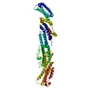
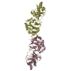
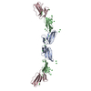
| Title | Crystal structure of the Brox Bro1 domain in complex with the C-terminal tail of CHMP5 | ||||||
 Components Components |
| ||||||
 Keywords Keywords |  MEMBRANE PROTEIN/TRANSPORT PROTEIN / beta hairpin / ESCRT-III / CHMPs / MEMBRANE PROTEIN-TRANSPORT PROTEIN complex / BROX MEMBRANE PROTEIN/TRANSPORT PROTEIN / beta hairpin / ESCRT-III / CHMPs / MEMBRANE PROTEIN-TRANSPORT PROTEIN complex / BROX | ||||||
| Function / homology |  Function and homology information Function and homology informationamphisome membrane / multivesicular body-lysosome fusion / viral budding / vesicle fusion with vacuole / ESCRT III complex disassembly / late endosome to lysosome transport / kinetochore microtubule / late endosome to vacuole transport via multivesicular body sorting pathway / mitotic nuclear membrane reassembly / regulation of centrosome duplication ...amphisome membrane / multivesicular body-lysosome fusion / viral budding / vesicle fusion with vacuole / ESCRT III complex disassembly / late endosome to lysosome transport / kinetochore microtubule / late endosome to vacuole transport via multivesicular body sorting pathway / mitotic nuclear membrane reassembly / regulation of centrosome duplication / cellular response to muramyl dipeptide / nuclear membrane reassembly / midbody abscission / multivesicular body sorting pathway / vesicle budding from membrane / membrane fission / plasma membrane repair / multivesicular body membrane / ubiquitin-dependent protein catabolic process via the multivesicular body sorting pathway / multivesicular body assembly / regulation of mitotic spindle assembly / mitotic metaphase chromosome alignment / nucleus organization / viral budding via host ESCRT complex / autophagosome membrane / autophagosome maturation / regulation of receptor recycling / nuclear pore / Endosomal Sorting Complex Required For Transport (ESCRT) / multivesicular body / erythrocyte differentiation / viral budding from plasma membrane / Budding and maturation of HIV virion / kinetochore / autophagy / protein transport / nuclear envelope / midbody / nuclear membrane / cellular response to lipopolysaccharide / cadherin binding / lysosomal membrane / extracellular exosome / plasma membrane / cytosolSimilarity search - Function | ||||||
| Biological species |  Homo sapiens (human) Homo sapiens (human) | ||||||
| Method | X-RAY DIFFRACTION / SYNCHROTRON / MOLECULAR REPLACEMENT / Resolution: 2.708 Å | ||||||
 Authors Authors | Jiang, J.S. / Mu, R.L. / Xiao, T. | ||||||
 Citation Citation | Journal: Structure / Year: 2012 Title: Two Distinct Binding Modes Define the Interaction of Brox with the C-Terminal Tails of CHMP5 and CHMP4B. Authors: Mu, R. / Dussupt, V. / Jiang, J. / Sette, P. / Rudd, V. / Chuenchor, W. / Bello, N.F. / Bouamr, F. / Xiao, T.S. | ||||||
| History |
|
- Structure visualization
| Structure viewer | Molecule: MolmilJmol/JSmol |
|---|
- Downloads & links
-Download
| PDBx/mmCIF format | 3um1.cif.gz | 168.6 KB | Display | PDBx/mmCIF format |
|---|---|---|---|---|
| PDB format | pdb3um1.ent.gz | 132.8 KB | Display | PDB format |
| PDBx/mmJSON format | 3um1.json.gz | Tree view | PDBx/mmJSON format | |
| Others |  Other downloads Other downloads |
-Validation report
| Arichive directory | https://data.pdbj.org/pub/pdb/validation_reports/um/3um1ftp://data.pdbj.org/pub/pdb/validation_reports/um/3um1 | HTTPS FTP |
|---|
-Related structure data
| Related structure data |  3ulyC  3um0C  3um2C  3um3C  3r9mS S: Starting model for refinement C: citing same article ( |
|---|---|
| Similar structure data |


-Links
 PDBj
PDBj

- Assembly
| Deposited unit | 
| ||||||||
|---|---|---|---|---|---|---|---|---|---|
| 1 | 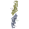
| ||||||||
| Unit cell |
|
-Components
| #1: Protein | Mass: 42533.410 Da / Num. of mol.: 2 / Fragment: Brox bro1 domain 2-377 Source method: isolated from a genetically manipulated source Source: (gene. exp.) Homo sapiens (human) / Gene: BROFTI, BROX, C1orf58 / Plasmid: pET30a / Production host:  Escherichia coli (E. coli) / Strain (production host): BL21(DE3) / References: UniProt: Q5VW32 Escherichia coli (E. coli) / Strain (production host): BL21(DE3) / References: UniProt: Q5VW32#2: Protein | Mass: 7294.708 Da / Num. of mol.: 2 / Fragment: C-terminal tail of CHMP5 151-219 Source method: isolated from a genetically manipulated source Source: (gene. exp.) Homo sapiens (human)Gene: C9orf83, CGI-34, CHMP5, HSPC177, PNAS-114, PNAS-2, SNF7DC2 Plasmid: pET30a / Production host: Escherichia coli (E. coli) / Strain (production host): BL21(DE3) / References: UniProt: Q9NZZ3#3: Chemical | ChemComp-GOL / | Glycerol  Mass: 92.094 Da / Num. of mol.: 1 / Source method: obtained synthetically / Formula: C3H8O3 Mass: 92.094 Da / Num. of mol.: 1 / Source method: obtained synthetically / Formula: C3H8O3#4: Water | ChemComp-HOH / | Water Mass: 18.015 Da / Num. of mol.: 157 / Source method: isolated from a natural source / Formula: H2O Mass: 18.015 Da / Num. of mol.: 157 / Source method: isolated from a natural source / Formula: H2O |
|---|
-Experimental details
-Experiment
| Experiment | Method: X-RAY DIFFRACTION / Number of used crystals: 1 |
|---|
- Sample preparation
| Crystal | Density Matthews: 2.24 Å3/Da / Density % sol: 45.07 % |
|---|---|
| Crystal grow | Temperature: 291 K / Method: vapor diffusion, hanging drop / pH: 6.5 Details: 20% PEG 8000, 0.1M sodium cacodylate, 0.2M magnesium acetate , pH 6.5, VAPOR DIFFUSION, HANGING DROP, temperature 291K |
-Data collection
| Diffraction | Mean temperature: 100 K |
|---|---|
| Diffraction source | Source: SYNCHROTRON / Site: NSLS  / Beamline: X29A / Wavelength: 1.075 Å / Beamline: X29A / Wavelength: 1.075 Å |
| Detector | Type: ADSC QUANTUM 315 / Detector: CCD / Date: Sep 3, 2011 |
| Radiation | Protocol: SINGLE WAVELENGTH / Monochromatic (M) / Laue (L): M / Scattering type: x-ray |
| Radiation wavelength | Wavelength: 1.075 Å / Relative weight: 1 |
| Reflection | Resolution: 2.7→50 Å / Num. obs: 23032 / % possible obs: 91.6 % / Observed criterion σ(F): 0 / Observed criterion σ(I): 0 / Redundancy: 4.3 % / Biso Wilson estimate: 38.1 Å2 / Rmerge(I) obs: 0.133 / Net I/σ(I): 7.1 |
- Processing
| Software |
| |||||||||||||||||||||||||||||||||||||||||||||||||||||||||||||||
|---|---|---|---|---|---|---|---|---|---|---|---|---|---|---|---|---|---|---|---|---|---|---|---|---|---|---|---|---|---|---|---|---|---|---|---|---|---|---|---|---|---|---|---|---|---|---|---|---|---|---|---|---|---|---|---|---|---|---|---|---|---|---|---|---|
| Refinement | Method to determine structure: MOLECULAR REPLACEMENT Starting model: PDB ENTRY 3R9M Resolution: 2.708→43.319 Å / SU ML: 0.36 / Cross valid method: THROUGHOUT / σ(F): 0 / Phase error: 25.45 / Stereochemistry target values: ML
| |||||||||||||||||||||||||||||||||||||||||||||||||||||||||||||||
| Solvent computation | Shrinkage radii: 0.95 Å / VDW probe radii: 1.2 Å / Solvent model: FLAT BULK SOLVENT MODEL / Bsol: 32.133 Å2 / ksol: 0.342 e/Å3 | |||||||||||||||||||||||||||||||||||||||||||||||||||||||||||||||
| Displacement parameters | Biso mean: 41.9 Å2
| |||||||||||||||||||||||||||||||||||||||||||||||||||||||||||||||
| Refinement step | Cycle: LAST / Resolution: 2.708→43.319 Å
| |||||||||||||||||||||||||||||||||||||||||||||||||||||||||||||||
| Refine LS restraints |
| |||||||||||||||||||||||||||||||||||||||||||||||||||||||||||||||
| LS refinement shell |
|
