National Institutes of Health/National Institute of General Medical Sciences (NIH/NIGMS)
GM080532
United States
Citation
Journal: To Be Published Title: PAK2 Phosphorylation Inhibits Caspase-7 by Two Divergent Mechanisms: Slowing Activation and Blocking Substrate Binding Authors: Eron, S.J. / Hardy, J.A.
Evidence: gel filtration, mass spectrometry, native gel electrophoresis
Type
Name
Symmetry operation
Number
identity operation
1_555
x,y,z
1
Buried area
13170 Å2
ΔGint
-82 kcal/mol
Surface area
18680 Å2
Method
PISA
Unit cell
Length a, b, c (Å)
88.757, 88.757, 185.141
Angle α, β, γ (deg.)
90.000, 90.000, 120.000
Int Tables number
154
Space group name H-M
P3221
Noncrystallographic symmetry (NCS)
NCS domain:
ID
Ens-ID
Details
1
1
A
2
1
C
1
2
B
2
2
D
NCS domain segments:
Component-ID: 0 / Refine code: 0
Dom-ID
Ens-ID
Beg auth comp-ID
Beg label comp-ID
End auth comp-ID
End label comp-ID
Auth asym-ID
Label asym-ID
Auth seq-ID
Label seq-ID
1
1
THR
THR
GLN
GLN
A
A
57 - 196
57 - 196
2
1
THR
THR
GLN
GLN
C
C
357 - 496
57 - 196
1
2
LYS
LYS
SER
SER
B
B
212 - 302
14 - 104
2
2
LYS
LYS
SER
SER
D
D
512 - 602
14 - 104
NCS ensembles :
ID
1
2
Details
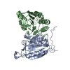
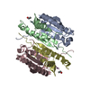
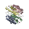
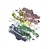
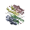
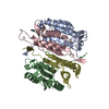
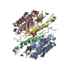
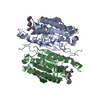
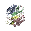
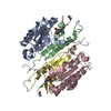
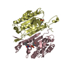
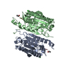
tetramer according to gel filtration, mass spectrometry, gel electroporesis, literature reports. This structure is of active, cleaved caspase-7. A procaspase-7 dimer becomes active, cleaved caspase-7 upon proteolytic processing, which cleaves after residues D23, D198 and D206. This generates four chains from each monomer. The prodomain (residues 1-23) and the intersubunit linker (residues 199-206) are both released. Thus active, cleaved caspase-7 has two copies of the large subunit (residues 24-198) and two copies of the small subunit (residues 206-303).
Mass: 22189.203 Da / Num. of mol.: 2 Source method: isolated from a genetically manipulated source Details: This structure is of active, cleaved caspase-7. A procaspase-7 dimer becomes active, cleaved caspase-7 upon proteolytic processing, which cleaves after residues D23, D198 and D206. This ...Details: This structure is of active, cleaved caspase-7. A procaspase-7 dimer becomes active, cleaved caspase-7 upon proteolytic processing, which cleaves after residues D23, D198 and D206. This generates four chains from each monomer. The prodomain (residues 1-23) and the intersubunit linker (residues 199-206) are both released. Thus active, cleaved caspase-7 has two copies of the large subunit (residues 24-198) and two copies of the small subunit (residues 206-303). The large subunit of caspase-7 crystallized here consisted of residues 24-198. Source: (gene. exp.) Homo sapiens (human) / Gene: CASP7, MCH3 / Production host: Escherichia coli (E. coli) / Strain (production host): BL21 (DE3) / References: UniProt: P55210, caspase-7
Mass: 13265.836 Da / Num. of mol.: 2 / Mutation: S272E Source method: isolated from a genetically manipulated source Details: This structure is of active, cleaved caspase-7. A procaspase-7 dimer becomes active, cleaved caspase-7 upon proteolytic processing, which cleaves after residues D23, D198 and D206. This ...Details: This structure is of active, cleaved caspase-7. A procaspase-7 dimer becomes active, cleaved caspase-7 upon proteolytic processing, which cleaves after residues D23, D198 and D206. This generates four chains from each monomer. The prodomain (residues 1-23) and the intersubunit linker (residues 199-206) are both released. Thus active, cleaved caspase-7 has two copies of the large subunit (residues 24-198) and two copies of the small subunit (residues 206-303). The small subunit of caspase-7 crystallized here consisted of residues 206-303 (including a 6xHis tag). Source: (gene. exp.) Homo sapiens (human) / Gene: CASP7, MCH3 / Production host: Escherichia coli (E. coli) / Strain (production host): BL21(DE3) / References: UniProt: P55210, caspase-7
Resolution: 2.2→76.98 Å / Cor.coef. Fo:Fc: 0.969 / Cor.coef. Fo:Fc free: 0.953 / SU B: 12.25 / SU ML: 0.139 / Cross valid method: THROUGHOUT / σ(F): 0 / ESU R: 0.174 / ESU R Free: 0.162 Details: HYDROGENS HAVE BEEN ADDED IN THE RIDING POSITIONS U VALUES: WITH TLS ADDED
Rfactor
Num. reflection
% reflection
Selection details
Rfree
0.2279
4298
9.9 %
RANDOM
Rwork
0.1912
-
-
-
obs
0.1948
39208
99.53 %
-
Solvent computation
Ion probe radii: 0.8 Å / Shrinkage radii: 0.8 Å / VDW probe radii: 1.2 Å
In the structure databanks used in Yorodumi, some data are registered as the other names, "COVID-19 virus" and "2019-nCoV". Here are the details of the virus and the list of structure data.
Jan 31, 2019. EMDB accession codes are about to change! (news from PDBe EMDB page)
EMDB accession codes are about to change! (news from PDBe EMDB page)
The allocation of 4 digits for EMDB accession codes will soon come to an end. Whilst these codes will remain in use, new EMDB accession codes will include an additional digit and will expand incrementally as the available range of codes is exhausted. The current 4-digit format prefixed with “EMD-” (i.e. EMD-XXXX) will advance to a 5-digit format (i.e. EMD-XXXXX), and so on. It is currently estimated that the 4-digit codes will be depleted around Spring 2019, at which point the 5-digit format will come into force.
The EM Navigator/Yorodumi systems omit the EMD- prefix.
Related info.:Q: What is EMD? / ID/Accession-code notation in Yorodumi/EM Navigator
Yorodumi is a browser for structure data from EMDB, PDB, SASBDB, etc.
This page is also the successor to EM Navigator detail page, and also detail information page/front-end page for Omokage search.
The word "yorodu" (or yorozu) is an old Japanese word meaning "ten thousand". "mi" (miru) is to see.
Related info.:EMDB / PDB / SASBDB / Comparison of 3 databanks / Yorodumi Search / Aug 31, 2016. New EM Navigator & Yorodumi / Yorodumi Papers / Jmol/JSmol / Function and homology information / Changes in new EM Navigator and Yorodumi
 Movie
Movie Controller


 Open data
 Basic information
 Components
 Keywords
 HYDROLASE /
  Function and homology information
 Authors
 United States, 1items
  Citation
 Structure visualization
 Downloads & links
 Other downloads
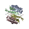


 PDBj
PDBj


 Assembly


 Mass: 46.025 Da / Num. of mol.: 3 / Source method: obtained synthetically / Formula: CH2O2
 Mass: 18.015 Da / Num. of mol.: 92 / Source method: isolated from a natural source / Formula: H2O
 Sample preparation
 Processing